Review
. 2012:2012:718617.
doi: 10.1155/2012/718617. Epub 2012 May 14.

Dextran sodium sulphate colitis mouse model: traps and tricks

Affiliations
Review

Dextran sodium sulphate colitis mouse model: traps and tricks

Martina Perše et al. J Biomed Biotechnol. 2012.

Abstract

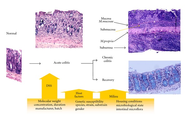
Inflammatory bowel disease (IBD) is a complex multifactorial disease of unknown etiology. Thus, dozens of different animal models of IBD have been developed in past decades. Animal models of IBD are valuable and indispensable tools that provide a wide range of options for investigating involvement of various factors into the pathogenesis of IBD and to evaluate different therapeutic options. However, the dextran sulphate sodium (DSS-) induced colitis model has some advantages when compared to other animal models of colitis. It is well appreciated and widely used model of inflammatory bowel disease because of its simplicity. It has many similarities to human IBD, which are mentioned in the paper. In spite of its simplicity and wide applicability, there are also traps that need to be taken into account when using DSS model. As demonstrated in the present paper, various factors may affect susceptibility to DSS-induced lesions and modify results.


Figures

Figure 1
Figure 1
Schematic simplified representation of various factors that can influence the susceptibility, onset, severity, and responsiveness to DSS-induced colitis.
Figure 2
Figure 2
(a) Disappearance of crypts. (b) Erosion and phlegmonous inflammation of mucosa and submucosa. Kreyberg trichrom stain (acid mucopolysaccharides are stained blue). C57BL/6JOlaHsd female mice are exposed to 3% DSS solution for 5 days followed by drinking water for 7 days.
Figure 3
Figure 3
(a) Vacuolar hydropic degeneration of cells. (b) Epithelial necrosis, cryptitis, and crypt abscesses. Kreyberg trichrom stain (acid mucopolysaccharides are stained blue). C57BL/6JOlaHsd female mice exposed to 3% DSS solution for 5 days followed by drinking water for 21 days.
Figure 4
Figure 4
Arrows denote crypt abscesses (neutrophils) in the lumen of crypts. Kreyberg trichrom stain (acid mucopolysaccharides are stained blue). C57BL/6JOlaHsd male mice are exposed to 3% DSS solution for 5 days followed by drinking water for (a) 7 days and (b) 28 days.
Figure 5
Figure 5
(a) Mononuclear leucocytes infiltration, crypt architectural disarray, and deep mucosal lymphocytosis. (b) Focally there is a moderate epithelial regeneratory atypia simulating dysplasia (arrow). Kreyberg trichrom stain (acid mucopolysaccharides are stained blue). C57BL/6JOlaHsd male mice are exposed to 3% DSS solution for 5 days followed by drinking water for 28 days.
Figure 6
Figure 6
(a) Focal transmural chronic colitis (skip lesion). C57BL/6JOlaHsd female mice are exposed to 3% DSS solution for 5 days followed by drinking water for 28 days. (b) Transmural inflammation with lymphoid follicles in subserosa (arrows) and chronic erosion. C57BL/6JOlaHsd female mice are exposed to 3% DSS solution for 9 days followed by drinking water for 28 days. Kreyberg trichrom stain (acid mucopolysaccharides are stained blue).
Figure 7
Figure 7
(a) and (b) reepithelisation of rectal and distal colonic erosion by squamous epithelium (arrows). C57BL/6JOlaHsd male mice are exposed to 3% DSS solution for 5 days followed by drinking water for 7 days. Kreyberg trichrom stain (acid mucopolysaccharides are stained blue).
Figure 8
Figure 8
Schematic representation of mucosal (colon) and systemic (spleen, MLN) immune responses in C57BL/6OlaHsd mice exposed to 3% DSS for 6 days followed by water for 19 days. Measurements were made on day 0, 1, 3, 5, 8, 12, and 25 as denoted by numbers. Schematic representation is based on results obtained by Hall et al. [23]. MLN: mesenteric lymph nodes, N: neutrophils, M: macrophages, DC: dendritic cells, T: T cells, B: B cells.

References

    1. Sartor RB, Muehlbauer M. Microbial host interactions in IBD: implications for pathogenesis and therapy. Current Gastroenterology Reports. 2007;9(6):497–507. - PubMed
    1. Sartor RB. Microbial influences in inflammatory bowel diseases. Gastroenterology. 2008;134(2):577–594. - PubMed
    1. Xavier RJ, Podolsky DK. Unravelling the pathogenesis of inflammatory bowel disease. Nature. 2007;448(7152):427–434. - PubMed
    1. Gismera CS, Aladren BS. Inflammatory bowel diseases: a disease (s) of modern times? Is incidence still increasing? World Journal of Gastroenterology. 2008;14(36):5491–5498. - PMC - PubMed
    1. Zhu LJ, Yang X, Yu XQ. Anti-TNF-alpha therapies in systemic lupus erythematosus. Journal of Biomedicine and Biotechnology. 2010;2010:8 pages. Article ID 465898. - PMC - PubMed

Publication types

Substances