Review
. 2012:2012:938138.
doi: 10.1100/2012/938138. Epub 2012 May 22.

Biology and biotechnology of follicle development

Affiliations
Review

Biology and biotechnology of follicle development

Gustavo Adolfo Palma et al. ScientificWorldJournal. 2012.

Abstract

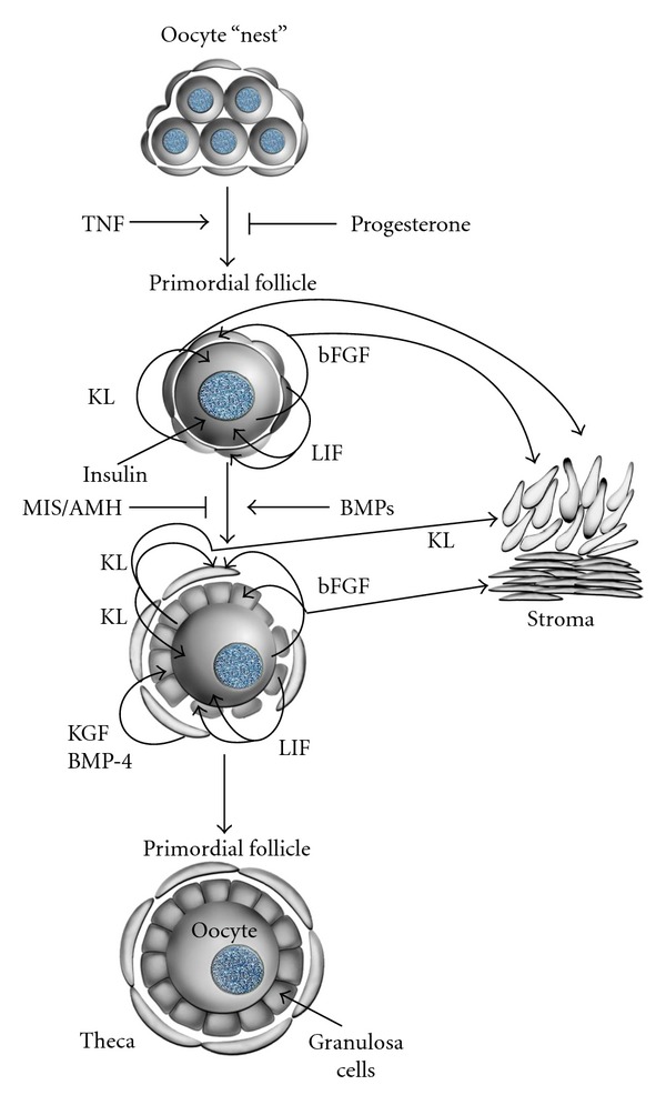
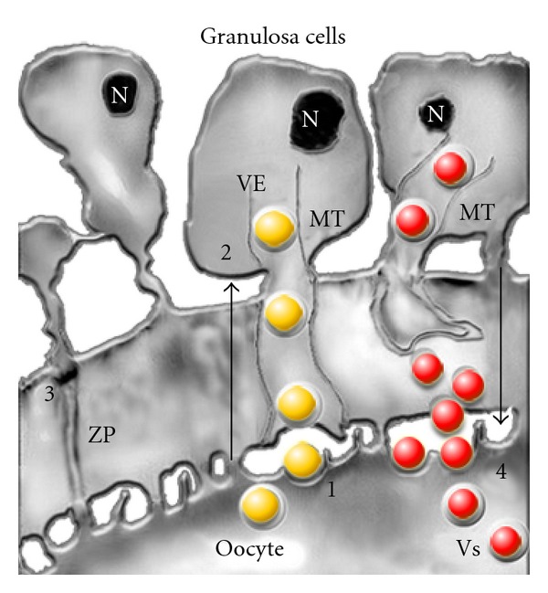
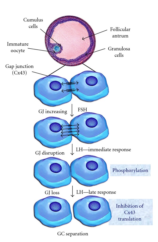
Growth and development of ovarian follicles require a series of coordinated events that induce morphological and functional changes within the follicle, leading to cell differentiation and oocyte development. The preantral early antral follicle transition is the stage of follicular development during which gonadotropin dependence is obtained and the progression into growing or atresia of the follicle is made. Follicular growth during this period is tightly regulated by oocyte-granulosatheca cell interactions. A cluster of early expressed genes is required for normal folliculogenesis. Granulosa cell factors stimulate the recruitment of theca cells from cortical stromal cells. Thecal factors promote granulosa cell proliferation and suppress granulosa cell apoptosis. Cell-cell and cell-extracellular matrix interactions influence the production of growth factors in the different follicular compartments (oocyte, granulosa, and theca cells). Several autocrine and paracrine factors are involved in follicular growth and differentiation; their activity is present even at the time of ovulation, decreasing the gap junction communication, and stimulating the theca cell proliferation. In addition, the identification of the factors that promote follicular growth from the preantral stage to the small antral stage may provide important information for the identification for assisted reproduction techniques.


Figures

Figure 1
Figure 1
Cell interactions in primordial follicles [3, 6]. Proposed cell interactions in the development of primordial follicles and the effect of the growth factors. Cell-cell interactions are mediated by tumor necrosis factor-alpha (TNFα), Kit ligand (KL), basic fibroblast growth (bFGF), leukemia inhibitory factor (LIF), bone morphogenetic protein-4 (BMP-4), keratinocyte growth factor (KGF), insulin, and anti-Mullerian hormone (AMH). With permission from M. Skinner.
Figure 2
Figure 2
Model proposing regulated delivery of the paracrine factors to the oocyte-granulosa cell interface [6, 7]. Four communication pathways are described. (1) Localized uptake of growth factors, like GDF-9, from the oocyte (yellow ball) by endocytosis at attachment sites of transzonal projections (TZPs) at the oolema. The vectorial transport of endocytic vesicles (VEs) to the cytoplasm of the granulosa cells takes place through the microtubules (MT). (2) Granulosa-zona pellucida interactions are necessary for the orientation of the TZPs. Contact sites may play an important role in the signaling role of oocytes and granulosa cells. (3) The gap junctions that allow direct intercellular communications between the microvilli of the oocyte and the granulosa cell TZPs. (4) Pathway for delivery of granulosa cell factors (red ball) packaged in secretory vesicles (VSs), which undergo endocytosis through the specific receptor on the oocyte surface. N: nucleus, MT: microtubules, ZP: zona pellucida, red ball: factors of the granulosa cells, yellow ball: factors released by the oocyte. With permission from D. F. Albertini.
Figure 3
Figure 3
Model of regulation of gap junctional communication (GJ, Cx43) [6, 8, 9] During development of the antral follicles FSH hormone stimulates mRNA expression that codifies the Cx43 synthesis of the GJs, the amplification of functional channels, and consequently the integration to the metabolic activity [8]. Mediated by the family mitogen-activated protein kinases, preovulatory LH levels interrupt cell-cell communications by means of phosphorylation and modification of Cx43 protein conformation. This leads to interruption of the intercellular channels. The primary effect of the immediate response to LH is accompanied by elimination of the Cx43 protein, disappearance of GJ sand separation of the GCs from the oocyte. GJ: gap junction; GC: granulosa cells. With permission from N. Dekel.
Figure 4
Figure 4
See [6, 10]. (a) Diameters of primordial follicles and primary follicles of 4 species. (b) Oocyte diameters in primordial follicles and primary oocytes of 4 species.
Figure 5
Figure 5
See [6, 10]. (a) Diameters of early antral follicles, and Graafian follicles of 4 species. (b) Oocyte diameters of early antral follicles and Graafian follicles of 4 species.
Figure 6
Figure 6
Morphology changes [6, 11, 12] Structure, function and change in the morphology of the granulosa cells [11], components of the basal lamina and the Call-Exner bodies of. (a) Primordial follicles and (b) preantral follicles [12]. With permission from Rodgers [11].
Figure 7
Figure 7
Model of a putative transcription network of gene, protein and hormonal stimulus of follicle development [6, 13, 14]. The process starts with the formation of germ cells and continues with the development of oogonia and oocytes. Several factors are involved in the PGC formation, oogonial (Oct-4), and oocyte development (Figα, Nobox, etc.). Specific oocyte differentiation factors in preantral follicular stages are, YP30, ZP1-3 and those of cortical granulosa content: DF1,9 ovoperoxidase (Opo), proteoliasin (Pln), and rendivin (Rdv). PMFs: Primordial follicles, PF: primary follicle, SF: secondary follicle, IGF (family of the insulin-like growth factors). With permission from Song and Wessel [14].

Similar articles

Cited by

References

    1. Ireland JJ, Zielak-Steciwko AE, Jimenez-Krassel F, et al. Variation in the ovarian reserve is linked to alterations in intrafollicular estradiol production and ovarian biomarkers of follicular differentiation and oocyte quality in cattle. Biology of Reproduction. 2009;80(5):954–964. - PubMed
    1. Fortune JE. The early stages of follicular development: Activation of primordial follicles and growth of preantral follicles. Animal Reproduction Science. 2003;78(3-4):135–163. - PubMed
    1. Skinner MK. Regulation of primordial follicle assembly and development. Human Reproduction Update. 2005;11(5):461–471. - PubMed
    1. McNatty KP, Fidler AE, Juengel JL, et al. Growth and paracrine factors regulating follicular formation and cellular function. Molecular and Cellular Endocrinology. 2000;163(1-2):11–20. - PubMed
    1. Hyttel P, Sinowatz F, Vejlsted M. Domestic Animal Embryology. Edinburgh, UK: Saunders Elsevier; 2010.

LinkOut - more resources