. 2012:2012:219625.
doi: 10.1155/2012/219625. Epub 2012 May 15.

Autophagy: more than a nonselective pathway

Affiliations

Autophagy: more than a nonselective pathway

Fulvio Reggiori et al. Int J Cell Biol. 2012.

Abstract

Autophagy is a catabolic pathway conserved among eukaryotes that allows cells to rapidly eliminate large unwanted structures such as aberrant protein aggregates, superfluous or damaged organelles, and invading pathogens. The hallmark of this transport pathway is the sequestration of the cargoes that have to be degraded in the lysosomes by double-membrane vesicles called autophagosomes. The key actors mediating the biogenesis of these carriers are the autophagy-related genes (ATGs). For a long time, it was assumed that autophagy is a bulk process. Recent studies, however, have highlighted the capacity of this pathway to exclusively eliminate specific structures and thus better fulfil the catabolic necessities of the cell. We are just starting to unveil the regulation and mechanism of these selective types of autophagy, but what it is already clearly emerging is that structures targeted to destruction are accurately enwrapped by autophagosomes through the action of specific receptors and adaptors. In this paper, we will briefly discuss the impact that the selective types of autophagy have had on our understanding of autophagy.


Figures

Figure 1
Figure 1
Multiple Atg proteins govern autophagosome formation. In response to inactivation of mTORC1 (but also other cellular and environmental cues), the ULK1 complex is activated and translocates in proximity of the endoplasmic reticulum (ER). Thereafter, the ULK1 complex regulates the class III PI3K complex. Atg9L, a multimembrane spanning protein, is also involved in an early stage of autophagosome formation by probably supplying part of the membranes necessary for the formation and/or expansion. Local formation of PI3P at sites called omegasomes promotes the formation of the phagophore, from which autophagosomes appear to be generated. The PI3P-binding WIPI proteins (yeast Atg18 homolog), as well as the Atg12-Atg5-Atg16L1 complex and the LC3-phosphatidylethanolamine (PE) conjugate play important roles in the elongation and closure of the isolation membrane. Finally, the complete autophagosome fuses with endosomes or endosome-derived vesicles forming the amphisome, which subsequently fuses with lysosomes to form autolysosomes. In the lysosomes, the cytoplasmic materials engulfed by the autophagosomes are degraded by resident hydrolases. The resulting amino acids and other basic cellular constituents are reused by the cell; when in high levels they also reactivate mTORC1 and then suppress autophagy.
Figure 2
Figure 2
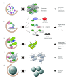
Representative selective autophagy. (a) The cytoplasm-to-vacuole targeting (Cvt) pathway. Ape1 is synthesized as a cytoplasmic precursor protein with a propeptide and rapidly oligomerizes into dodecamers that subsequently associate with each other to form a higher order complex. The autophagy receptor Atg19 directly binds to the complex and mediates the recruitment of another Cvt pathway cargo, Ams1, leading to the formation of the so-called Cvt complex. Atg19 also interacts with the autophagy adaptor Atg11 and this protein allows the transport of the Cvt complex to the site where the double-membrane vesicle will be generated. At this location, Atg11 tethers the Atg proteins essential for the Cvt vesicle formation and the direct binding of Atg19 to Atg8 permits the exclusive sequestration of the Cvt complex into the vesicle. (b) A model for p62 and NBR1 as autophagy receptors for ubiquitinated cargos. p62 and NBR1 bind with ubiquitinated cargos via their ubiquitin-associated (UBA) domain and this interaction triggers the aggregate formation through the oligomerization of p62 via its Phox and Bem1p (PB1) domain. Furthermore, p62 interacts with both autophagy-linked FYVE protein (ALFY), which serves to recruit Atg5 and to bind PI3P, and directly with LC3. This latter event appears to organize and activate the Atg machinery in close proximity of the ubiquitinated cargos, which allows to selectively sequester them in the autophagosomes in analogous to the Cvt pathway.
Figure 3
Figure 3
Pathophysiological relevance of selective autophagy. (a, b) Selective types of autophagy operates constitutively at low levels even under nutrient-rich conditions and mediates turnover of selected cytoplasmic materials through the action of autophagy receptors such as p62 and NBR1. These proteins mediate the elimination of ubiquitinated structures, including protein aggregates (a) and defects in these pathways lead to the disruption of tissue homeostasis, resulting in life-threatening diseases. Defective autophagy is usually accompanied by extensive accumulation of p62-containing aggregates, which enhances its function as a scaffold protein in several signaling cascades such as NF-κB signaling, apoptosis, and Nrf2 activation (b). Such abnormalities might be involved in tumorigenesis and Paget's disease of bone. (c) During erythroid differentiation, Nix/Bnip3L relocalization to mitochondria leads to their depolarization, which triggers mitophagy. Loss of Nix/Bnip3L causes an arrest in the erythroid maturation arrest, leading to severe anaemia. In response to loss of the mitochondrial membrane potential, Parkin translocates onto the damaged mitochondria in a PINK1-dependent manner, and ubiquitinated proteins present on the outer mitochondrial membrane, which induces mitophagy. Parkinson's disease-related mutations in the Parkin and PINK1 genes provoke a defect in mitophagy, suggesting this selective type of autophagy has a role in preventing the pathogenesis of the Parkinson's disease. (d) Specific bacteria invading the cytosol get ubiquitinated and are recognized by autophagy receptors such as p62, NDP52, and optineurin (OPTN). This allows the specific sequestration of the microbes into autophagosomes and their delivery into the lysosomes for degradation. (e) The lipid droplets are probably degraded by autophagy selectively. This selective type of autophagy, lipophagy, supplies free-fatty acids utilized to generate energy through the β-oxidation. Impairments in lipophagy are known to cause accumulation of lipid droplets in hepatocytes and reduced production of AgRP in neurons.

References

    1. Arias E, Cuervo AM. Chaperone-mediated autophagy in protein quality control. Current Opinion in Cell Biology. 2011;23(2):184–189. - PMC - PubMed
    1. Orenstein SJ, Cuervo AM. Chaperone-mediated autophagy: molecular mechanisms and physiological relevance. Seminars in Cell and Developmental Biology. 2010;21(7):719–726. - PMC - PubMed
    1. Mijaljica D, Prescott M, Devenish RJ. Microautophagy in mammalian cells: revisiting a 40-year-old conundrum. Autophagy. 2011;7(7):673–682. - PubMed
    1. Sahu R, Kaushik S, Clement CC, et al. Microautophagy of cytosolic proteins by late endosomes. Developmental Cell. 2011;20(1):131–139. - PMC - PubMed
    1. Eskelinen E-L, Reggiori F, Baba M, Kovács AL, Seglen PO. Seeing is believing: the impact of electron microscopy on autophagy research. Autophagy. 2011;7(9):935–956. - PubMed

LinkOut - more resources