

Retinal axonal loss begins early in the course of multiple sclerosis and is similar between progressive phenotypes
- PMID: 22666330
- PMCID: PMC3359324
- DOI: 10.1371/journal.pone.0036847
Retinal axonal loss begins early in the course of multiple sclerosis and is similar between progressive phenotypes
Abstract
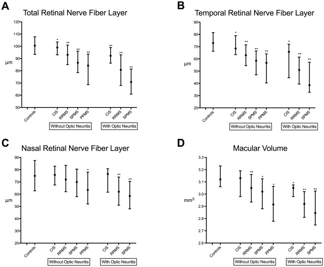
Background: To determine whether retinal axonal loss is detectable in patients with a clinically isolated syndrome (CIS), a first clinical demyelinating attack suggestive of multiple sclerosis (MS), and examine patterns of retinal axonal loss across MS disease subtypes.
Methodology/principal findings: Spectral-domain Optical Coherence Tomography was performed in 541 patients with MS, including 45 with high-risk CIS, 403 with relapsing-remitting (RR)MS, 60 with secondary-progressive (SP)MS and 33 with primary-progressive (PP)MS, and 53 unaffected controls. Differences in retinal nerve fiber layer (RNFL) thickness and macular volume were analyzed using multiple linear regression and associations with age and disease duration were examined in a cross-sectional analysis. In eyes without a clinical history of optic neuritis (designated as "eyes without optic neuritis"), the total and temporal peripapillary RNFL was thinner in CIS patients compared to controls (temporal RNFL by -5.4 µm [95% CI -0.9 to--9.9 µm, p = 0.02] adjusting for age and sex). The total (p = 0.01) and temporal (p = 0.03) RNFL was also thinner in CIS patients with clinical disease for less than 1 year compared to controls. In eyes without optic neuritis, total and temporal RNFL thickness was nearly identical between primary and secondary progressive MS, but total macular volume was slightly lower in the primary progressive group (p<0.05).
Conclusions/significance: Retinal axonal loss is increasingly prominent in more advanced stages of disease--progressive MS>RRMS>CIS--with proportionally greater thinning in eyes previously affected by clinically evident optic neuritis. Retinal axonal loss begins early in the course of MS. In the absence of clinically evident optic neuritis, RNFL thinning is nearly identical between progressive MS subtypes.
Conflict of interest statement
Figures

References
-
- Trapp BD, Peterson J, Ransohoff RM, Rudick R, Mork S, et al. Axonal transection in the lesions of multiple sclerosis. N Engl J Med. 1998;338:278–285. - PubMed
-
- Bitsch A, Schuchardt J, Bunkowski S, Kuhlmann T, Bruck W. Acute axonal injury in multiple sclerosis. Correlation with demyelination and inflammation. Brain. 2000;123(Pt 6):1174–1183. - PubMed
-
- Fisher E, Lee JC, Nakamura K, Rudick RA. Gray matter atrophy in multiple sclerosis: a longitudinal study. Ann Neurol. 2008;64:255–265. - PubMed
-
- Audoin B, Zaaraoui W, Reuter F, Rico A, Malikova I, et al. Atrophy mainly affects the limbic system and the deep grey matter at the first stage of multiple sclerosis. J Neurol Neurosurg Psychiatry. 2010;81:690–695. - PubMed
Publication types
MeSH terms
Grants and funding
LinkOut - more resources
Full Text Sources
Medical
Miscellaneous
