. 2012;7(5):e37222.
doi: 10.1371/journal.pone.0037222. Epub 2012 May 30.

Dihydroartemisinin enhances Apo2L/TRAIL-mediated apoptosis in pancreatic cancer cells via ROS-mediated up-regulation of death receptor 5

Affiliations

Dihydroartemisinin enhances Apo2L/TRAIL-mediated apoptosis in pancreatic cancer cells via ROS-mediated up-regulation of death receptor 5

Rui Kong et al. PLoS One. 2012.

Erratum in

  • PLoS One. 2012;7(10). doi:10.1371/annotation/f7203563-87dc-4d11-a1b7-958f81cf743a

Abstract

Background: Dihydroartemisinin (DHA), a semi-synthetic derivative of artemisinin, has recently shown antitumor activity in various cancer cells. Apo2 ligand or tumor necrosis factor-related apoptosis-inducing ligand (Apo2L/TRAIL) is regarded as a promising anticancer agent, but chemoresistance affects its efficacy as a treatment strategy. Apoptosis induced by the combination of DHA and Apo2L/TRAIL has not been well documented, and the mechanisms involved remain unclear.

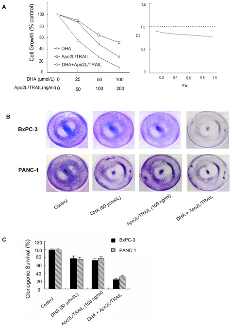
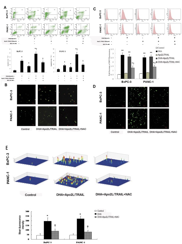
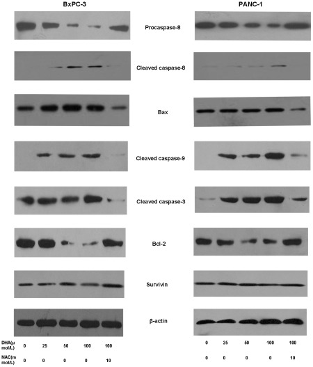
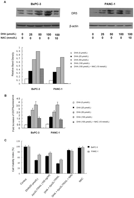
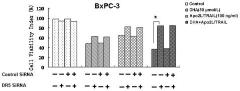
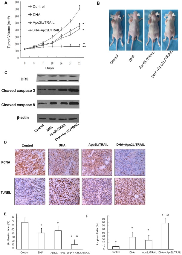
Methodology/principal findings: Here, we report that DHA enhances the efficacy of Apo2L/TRAIL for the treatment of pancreatic cancer. We found that combined therapy using DHA and Apo2L/TRAIL significantly enhanced apoptosis in BxPC-3 and PANC-1 cells compared with single-agent treatment in vitro. The effect of DHA was mediated through the generation of reactive oxygen species, the induction of death receptor 5 (DR5) and the modulation of apoptosis-related proteins. However, N-acetyl cysteine significantly reduced the enhanced apoptosis observed with the combination of DHA and Apo2L/TRAIL. In addition, knockdown of DR5 by small interfering RNA also significantly reduced the amount of apoptosis induced by DHA and Apo2L/TRAIL.

Conclusions/significance: These results suggest that DHA enhances Apo2L/TRAIL-mediated apoptosis in human pancreatic cancer cells through reactive oxygen species-mediated up-regulation of DR5.


Conflict of interest statement

Competing Interests: The authors have declared that no competing interests exist.

Figures

Figure 1
Figure 1. DHA synergistically enhances Apo2L/TRAIL-induced cell death in BxPC-3 cells.
(A) Cells were treated with DHA alone, Apo2L/TRAIL alone or a combination of the two agents and incubated at 37°C for 72 h. The viability of the cells was assessed using the MTT method. Combination index (CI) versus fraction affected (Fa) plots obtained from median-effect analysis of Chou-Talalay. A CI>1 indicates antagonism,  = 1 indicates additivity, and <1 indicates synergy. (B) The clonogenic assay was performed as described in the Materials and Methods section. The cells were treated with DHA (50 µmol/L) alone, Apo2L/TRAIL (100 ng/ml) alone or the combination of the two drugs for 24 h and washed with PBS. The cells were then incubated for an additional 7 d and stained with crystal violet. (C) Clonogenic survival is presented as the percentage of surviving colonies formed in drug-treated cells with respect to untreated cells.
Figure 2
Figure 2. DHA-induced apoptosis is caused by ROS.
(A) Apoptosis of pancreatic cancer cells. BxPC-3 and PANC-1 cells were treated with DHA (50 µmol/L) and Apo2L/TRAIL (100 ng/ml) as indicated. Flow cytometry was performed to measure apoptosis rates (%). A significant increase in the apoptosis rate compared with the control is denoted by “*” (P<0.05), a significant increase compared with DHA- or Apo2L/TRAIL-treated cells is denoted by “†” (P<0.01), and a significant decrease compared with DHA+Apo2L/TRAIL-treated cells is denoted by “‡” (P<0.01). Representative histograms from cytometrically analyzed BxPC-3 and PANC-1 cells treated with control, DHA, Apo2L/TRAIL, DHA+Apo2L/TRAIL or NAC (10 mM). (B) Laser scanning confocal microscopy of cells. Representative photographs were taken of the control BxPC-3 and PANC-1 cells and of BxPC-3 and PANC-1 cells treated with DHA+Apo2L/TRAIL and DHA+Apo2L/TRAIL+NAC. (C) Levels of intracellular ROS measured in vitro. BxPC-3 and PANC-1 cells were treated with DHA (50 µmol/L), Apo2L/TRAIL (100 ng/ml), DHA+Apo2L/TRAIL, or pretreated with NAC (10 mM) and then treated with DHA+Apo2L/TRAIL for 6 h. Untreated cells served as the control. The cells were incubated with DCFHDA and then subjected to flow cytometry to measure the levels of intracellular ROS, as represented by DCF fluorescence. A significant increase in DCF fluorescence compared with the control is denoted by “*” (P<0.05), a highly significant difference compared with the control is denoted by “**” (P<0.01), and a significant reduction compared with the DHA+Apo2L/TRAIL treatment is denoted by “†” (P<0.05). (D) Representative photographs are shown for DCFHDA-stained cells observed using laser scanning confocal microscopy. The green fluorescence represents intracellular ROS. (E) The mean fluorescence intensity was measured for the DCFHDA-stained cells, and the respective 3-dimensional horizontal plane images were produced by laser scanning confocal microscopy. A significant difference from the control is denoted by “*” (P<0.01), and a significant difference from the DHA+Apo2L/TRAIL treatment is denoted by “†” (P<0.05).
Figure 3
Figure 3. The expression of apoptosis-related genes.
BxPC-3 and PANC-1 cells were treated with various concentrations of DHA (0, 25, 50, 100 µmol/L) and pretreated with NAC followed by DHA (100 µmol/L) for 72 h. Whole cell extracts were prepared and analyzed by western blotting using antibodies against Bcl-2, Bax, surviving, caspase-3, caspase-8, and caspase-9. DHA significantly up-regulated the expression of Bax, caspase-3, caspase-8 and caspase-9, and down-regulated the expression of Bcl-2. However, DHA had little influence on the expression of survivin. DHA with NAC (10 mM) pretreatment did not up-regulate the expression of caspase-8, Bax, caspase-9, and caspase-3. β-actin served as an internal control.
Figure 4
Figure 4. Up-regulation of DR5 by DHA was mediated by ROS.
(A) BxPC-3 and PANC-1 cells were treated with the indicated doses of DHA for 48 h. Whole cell extracts were prepared and analyzed for DR5 expression using western blotting. DHA had dose-dependent effects on the expression of DR5. The DHA-induced increase in DR5 levels was significantly blocked by pretreatment with 10 mM NAC. β-actin served as an internal control. (B) The cells were collected and analyzed using flow cytometry. A gradual increase in fluorescence was observed in cells treated with 25, 50 and 100 µmol/L DHA, respectively, indicating a dose-dependent increase in the production of ROS in response to DHA treatment in the two cell lines. The production of ROS was markedly inhibited by pretreating the cells with NAC (10 mM). (C) The cells were treated with DHA (50 µmol/L) alone, Apo2L/TRAIL (100 ng/ml) alone or a combination of the two agents. The cells were also treated with NAC (10 mM) alone or pretreated with NAC and incubated at 37°C for 72 h. The viability of the cells was assessed using the MTT method and the viability index (%) was calculated. Significant differences are denoted by “*” (P<0.01).
Figure 5
Figure 5. Effects of knockdown of DR5 expression on DHA-induced cytotoxicity and cell apoptosis of Apo2L/TRAIL.
BxPC-3 and PANC-1 cells were transfected with DR5 siRNA and control siRNA, either alone or in combination. After 48 h, the cells were treated with 50 µmol/L DHA for 24 h, and whole cell extracts were subjected to western blotting to test for the expression of DR5. Transfection of cells with siRNA targeting DR5 specifically silenced the expression of DR5. The cells were seeded on a chamber slide and transfected with siRNAs. After 48 h, the cells were treated with 50 µmol/L DHA, 100 ng/mL Apo2L/TRAIL, either alone or in combination, and incubated at 37°C for 72 h. The viability of the cells was assessed using the MTT method, and the viability index (%) was calculated. Silencing of DR5 by siRNA reduced the cytotoxic effect of the combination of DHA and Apo2L/TRAIL but not of DHA alone. Significant differences are denoted by “*” (P<0.01).
Figure 6
Figure 6. Tumor growth, tumor gene expression, tumor proliferation and apoptosis in vivo.
(A) BxPC-3 tumors were established subcutaneously in mice. When the tumors reached approximately 120 mm3 in volume, the mice were randomly assigned to control, DHA, Apo2L/TRAIL, or DHA+Apo2L/TRAIL groups and treated as described in the methods section. The sizes (measured in mm3) of the tumors were monitored and recorded. A significant difference in tumor volume from the control is denoted by “*” (P<0.05), and a significant reduction compared to the DHA or Apo2L/TRAIL-treated tumors is denoted by “**” (P<0.01). (B) Representative animals and tumors are shown for each group. (C) Tumors from control mice and from mice treated with DHA, Apo2L/TRAIL, and DHA+Apo2L/TRAIL were homogenized and subjected to western blot analysis to detect the expression of caspase-3 and caspase-8. β-actin served as an internal control. (D) Analysis of proliferation marker PCNA by immunohistochemistry and apoptotic status of tumor cells by in situ TUNEL assay. PCNA positive (E) and TUNEL-positive (F) cells were also counted under microscope to calculate the proliferation index and apoptotic index, respectively. “*”: P<0.05, compared with control. “**”: P<0.01, compared with single agent.

Similar articles

Cited by

References

    1. Raraty MG, Magee CJ, Ghaneh P, Neoptolemos JP. New techniques and agents in the adjuvant therapy of pancreatic cancer. Acta Oncol. 2002;41:582–595. - PubMed
    1. Beger HG, Rau B, Gansauge F, Poch B, Link KH. Treatment of pancreatic cancer: Challenge of the facts. World J Surg. 2003;27:1075–1084. - PubMed
    1. Jemal A, Siegel R, Ward E, Hao Y, Xu J, et al. Cancer statistics, 2008. CA Cancer J Clin. 2008;58:71–96. - PubMed
    1. Meshnick SR. Artemisinin: mechanisms of action, resistance and toxicity. Int J Parasitol. 2002;32:1655–1660. - PubMed
    1. O'Neill PM, Posner GH. A medicinal chemistry perspective on artemisinin and related endoperoxides. J Med Chem. 2004;47:2945–2964. - PubMed

Publication types

MeSH terms

Substances