Comparative Study
. 2012;7(5):e37439.
doi: 10.1371/journal.pone.0037439. Epub 2012 May 29.

Citrulline a more suitable substrate than arginine to restore NO production and the microcirculation during endotoxemia

Affiliations
Comparative Study

Citrulline a more suitable substrate than arginine to restore NO production and the microcirculation during endotoxemia

Karolina A P Wijnands et al. PLoS One. 2012.

Abstract

Background: Impaired microcirculation during endotoxemia correlates with a disturbed arginine-nitric oxide (NO) metabolism and is associated with deteriorating organ function. Improving the organ perfusion in endotoxemia, as often seen in patients with severe infection or systemic inflammatory response syndrome (SIRS) is, therefore, an important therapeutic target. We hypothesized that supplementation of the arginine precursor citrulline rather than arginine would specifically increase eNOS-induced intracellular NO production and thereby improve the microcirculation during endotoxemia.

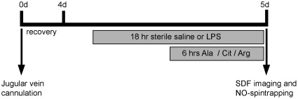
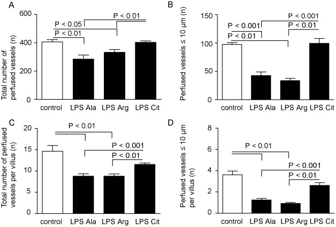
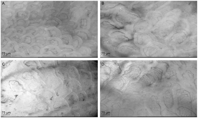
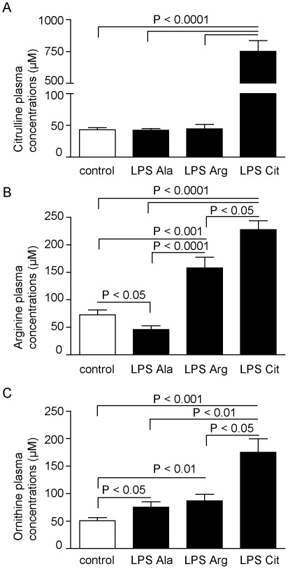
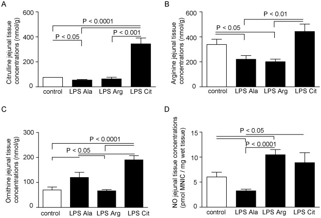
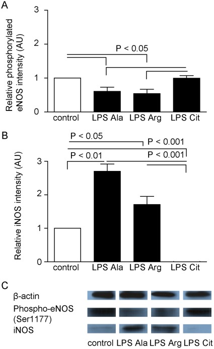
Methodology/principal findings: To study the effects of L-Citrulline and L-Arginine supplementation on jejunal microcirculation, intracellular arginine availability and NO production in a non-lethal prolonged endotoxemia model in mice. C57/Bl6 mice received an 18 hrs intravenous infusion of endotoxin (LPS, 0.4 µg • g bodyweight(-1) • h(-1)), combined with either L-Citrulline (6.25 mg • h-1), L-Arginine (6.25 mg • h(-1)), or L-Alanine (isonitrogenous control; 12.5 mg • h(-1)) during the last 6 hrs. The control group received an 18 hrs sterile saline infusion combined with L-Alanine or L-Citrulline during the last 6 hrs. The microcirculation was evaluated at the end of the infusion period using sidestream dark-field imaging of jejunal villi. Plasma and jejunal tissue amino-acid concentrations were measured by HPLC, NO tissue concentrations by electron-spin resonance spectroscopy and NOS protein concentrations using Western blot.

Conclusion/significance: L-Citrulline supplementation during endotoxemia positively influenced the intestinal microvascular perfusion compared to L-Arginine-supplemented and control endotoxemic mice. L-Citrulline supplementation increased plasma and tissue concentrations of arginine and citrulline, and restored intracellular NO production in the intestine. L-Arginine supplementation did not increase the intracellular arginine availability. Jejunal tissues in the L-Citrulline-supplemented group showed, compared to the endotoxemic and L-Arginine-supplemented endotoxemic group, an increase in degree of phosphorylation of eNOS (Ser 1177) and a decrease in iNOS protein level. In conclusion, L-Citrulline supplementation during endotoxemia and not L-Arginine reduced intestinal microcirculatory dysfunction and increased intracellular NO production, likely via increased intracellular citrulline and arginine availability.


Conflict of interest statement

Competing Interests: The authors recieved a grant from ESICM, the European society of Intensive Care medicine for our peer reviewed research proposal. Eli Lilly provided the price money to ESICM for this Sepsis Elite award in 2008 without any further conditions. This does not alter the authors' adherence to all the PloS ONE policies on sharing data and materials.

Figures

Figure 1
Figure 1. Experimental set up of the prolonged endotoxemia model.
Mice were fitted with a jugular vein cannula at t = 0 d. After 4 days (t = 4 d) an 18hours continuous infusion with lipopolysaccharides (LPS) was started, which was combined during the last 6 hours with an infusion of Citrulline (Cit), Arginine (Arg) or an isonitrogenous quantity of the placebo Alanine (Ala). After completing the treatment, sidestream dark-field (SDF) imaging was used to quantify the microcirculation in the jejunal villi or Nitric Oxide (NO) spin trapping with iron-diethyldithiocarbamate (DETC) complexes to measure NO production in vivo.
Figure 2
Figure 2. Circulation measurements with SDF imaging in the control, LPS-Ala, LPS-Arg and LPS-Cit groups.
(A) L-Citrulline supplementation improved the number (mean±SEM) of measurable perfused vessels in the jejunal microcirculation during endotoxemia, (B) in particular of vessels with a diameter ≤10 µm (P<0.01), compared to LPS-Ala and LPS-Arg supplemented animals. (C) The total number of perfused vessels per villus and (D) microcirculatory vessels with a diameter ≤10 µm per villus were also significantly lower in the LPS-Ala and LPS-Arg supplemented animals compared to the control and LPS-Cit group.
Figure 3
Figure 3. Representative live images of the microcirculatory measurements in jejunal villi with SDF imaging.
(A) Representative live image of the jejunal microcirculation in a control mouse. (B) Representative live image of the jejunal microcirculation in a LPS-Ala treated mouse which shows only perfusion of the larger vessels. (C) Representative live image of a LPS-Arg treated mouse which shows a comparable perfusion pattern as the LPS-Ala treated mouse. (D) Representative live image of a LPS-Cit treated mouse, which’s shows more small perfused vessels per villus.
Figure 4
Figure 4. Amino-acid concentrations in plasma.
(A) Citrulline plasma concentrations increased after L-Citrulline supplementation (LPS-Cit) compared to all other groups (P<0.0001). (B) Plasma arginine concentrations were significantly reduced in the LPS-Ala group compared to the control group (P<0.05), whereas this concentration was significantly increased by L-Arginine or L-Citrulline supplementation (LPS-Arg and LPS-Cit group) compared to both other groups (P<0.001). (C) Plasma ornithine levels increased in the LPS-Ala treated group compared to the control group (P<0.05) and increased further upon L-Arginine or L-Citrulline supplementation.
Figure 5
Figure 5. Amino-acid concentrations and NO production in murine jejunal tissue.
(A) L-Citrulline supplementation (LPS-Cit) increased tissue citrulline concentration compared to the LPS-Ala, LPS-Arg and control groups (P<0.001) (B) Arginine concentrations were significantly reduced in the LPS-Ala group compared to the control group (P<0.05). Interestingly, this concentration was not increased by L-Arginine supplementation (LPS-Arg), whereas this concentration was significantly increased by L-Citrulline supplementation (LPS-Cit group; P<0.01). (C) Ornithine levels increased in the LPS-Ala treated group compared to the control group (P<0.05) and increased further upon L-Citrulline supplementation (P<0.0001 relative to the control group and P<0.05 relative to the LPS group). (D) In the jejunum, prolonged endotoxemia resulted in a significant decrease in NO production (measured as pmol mono-nitrosyl-iron complexes (MNIC)/mg wet tissue) compared to control (P<0.05). NO production was significantly improved in the LPS-Arg and LPS-Cit group compared to the LPS group (P<0.0001 and P<0.05 respectively).
Figure 6
Figure 6. Phosphorylated eNOS and iNOS protein levels in murine jejunal tissue.
(A) The degree of phosphorylation of eNOS protein (expressed in arbitrary units, AU) was higher in the LPS-Cit group than in the LPS-Ala and LPS-Arg group (P<0.05). (B) The iNOS protein concentration changed in an opposite detection, with significantly lower levels in the control (P<0.05) and LPS-Cit group (P<0.01) than in the LPS-Ala and LPS-Arg group. (C) Representative examples of expression of phosphorylated eNOS (Ser 1177), iNOS and beta-actin by Western blot analysis.

Similar articles

Cited by

References

    1. Ince C. The microcirculation unveiled. Am J Respir Crit Care Med. 2002;166:1–2. - PubMed
    1. Boerma EC, Mathura KR, van der Voort PH, Spronk PE, Ince C. Quantifying bedside-derived imaging of microcirculatory abnormalities in septic patients: a prospective validation study. Crit Care. 2005;9:R601–606. - PMC - PubMed
    1. Crouser ED, Julian MW, Dorinsky PM. Ileal VO(2)-O(2) alterations induced by endotoxin correlate with severity of mitochondrial injury. Am J Respir Crit Care Med. 1999;160:1347–1353. - PubMed
    1. Kaul DK, Zhang X, Dasgupta T, Fabry ME. Arginine therapy of transgenic-knockout sickle mice improves microvascular function by reducing non-nitric oxide vasodilators, hemolysis, and oxidative stress. Am J Physiol Heart Circ Physiol. 2008;295:H39–47. - PMC - PubMed
    1. Krejci V, Hiltebrand LB, Erni D, Sigurdsson GH. Endothelin receptor antagonist bosentan improves microcirculatory blood flow in splanchnic organs in septic shock. Crit Care Med. 2003;31:203–210. - PubMed

Publication types

MeSH terms