

Translation of EEG spatial filters from resting to motor imagery using independent component analysis
- PMID: 22666377
- PMCID: PMC3362620
- DOI: 10.1371/journal.pone.0037665
Translation of EEG spatial filters from resting to motor imagery using independent component analysis
Abstract
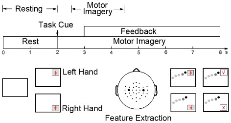
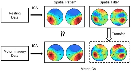
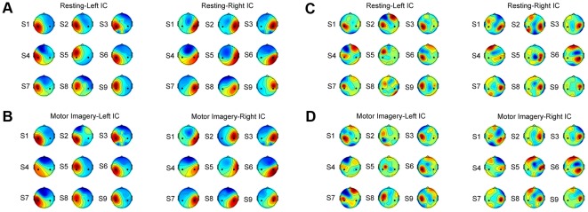
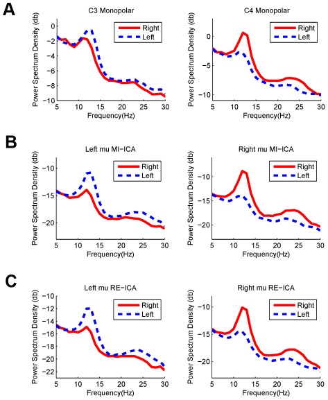
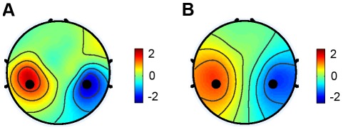
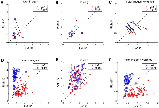
Electroencephalogram (EEG)-based brain-computer interfaces (BCIs) often use spatial filters to improve signal-to-noise ratio of task-related EEG activities. To obtain robust spatial filters, large amounts of labeled data, which are often expensive and labor-intensive to obtain, need to be collected in a training procedure before online BCI control. Several studies have recently developed zero-training methods using a session-to-session scenario in order to alleviate this problem. To our knowledge, a state-to-state translation, which applies spatial filters derived from one state to another, has never been reported. This study proposes a state-to-state, zero-training method to construct spatial filters for extracting EEG changes induced by motor imagery. Independent component analysis (ICA) was separately applied to the multi-channel EEG in the resting and the motor imagery states to obtain motor-related spatial filters. The resultant spatial filters were then applied to single-trial EEG to differentiate left- and right-hand imagery movements. On a motor imagery dataset collected from nine subjects, comparable classification accuracies were obtained by using ICA-based spatial filters derived from the two states (motor imagery: 87.0%, resting: 85.9%), which were both significantly higher than the accuracy achieved by using monopolar scalp EEG data (80.4%). The proposed method considerably increases the practicality of BCI systems in real-world environments because it is less sensitive to electrode misalignment across different sessions or days and does not require annotated pilot data to derive spatial filters.
Conflict of interest statement
Figures
 .
.
Similar articles
-
Generation of spatial filters by ICA for detecting motor-related oscillatory EEG.Annu Int Conf IEEE Eng Med Biol Soc. 2012;2012:1703-6. doi: 10.1109/EMBC.2012.6346276. Annu Int Conf IEEE Eng Med Biol Soc. 2012. PMID: 23366237
-
Motor Imagery EEG Classification Using Capsule Networks.Sensors (Basel). 2019 Jun 27;19(13):2854. doi: 10.3390/s19132854. Sensors (Basel). 2019. PMID: 31252557 Free PMC article.
-
A brain-computer interface driven by imagining different force loads on a single hand: an online feasibility study.J Neuroeng Rehabil. 2017 Sep 11;14(1):93. doi: 10.1186/s12984-017-0307-1. J Neuroeng Rehabil. 2017. PMID: 28893295 Free PMC article.
-
Improving the performance of an EEG-based motor imagery brain computer interface using task evoked changes in pupil diameter.PLoS One. 2015 Mar 27;10(3):e0121262. doi: 10.1371/journal.pone.0121262. eCollection 2015. PLoS One. 2015. PMID: 25816285 Free PMC article.
-
Diversity in a signal-to-image transformation approach for EEG-based motor imagery task classification.Med Biol Eng Comput. 2020 Feb;58(2):443-459. doi: 10.1007/s11517-019-02075-x. Epub 2019 Dec 21. Med Biol Eng Comput. 2020. PMID: 31863249
Cited by
-
A Fully Automated Trial Selection Method for Optimization of Motor Imagery Based Brain-Computer Interface.PLoS One. 2016 Sep 15;11(9):e0162657. doi: 10.1371/journal.pone.0162657. eCollection 2016. PLoS One. 2016. PMID: 27631789 Free PMC article.
-
Evolutionary Algorithm Based Feature Optimization for Multi-Channel EEG Classification.Front Neurosci. 2017 Feb 1;11:28. doi: 10.3389/fnins.2017.00028. eCollection 2017. Front Neurosci. 2017. PMID: 28203141 Free PMC article.
-
Utilizing sensory prediction errors for movement intention decoding: A new methodology.Sci Adv. 2018 May 9;4(5):eaaq0183. doi: 10.1126/sciadv.aaq0183. eCollection 2018 May. Sci Adv. 2018. PMID: 29750195 Free PMC article.
-
Aging Modulates the Resting Brain after a Memory Task: A Validation Study from Multivariate Models.Entropy (Basel). 2019 Apr 17;21(4):411. doi: 10.3390/e21040411. Entropy (Basel). 2019. PMID: 33267125 Free PMC article.
-
Review of brain encoding and decoding mechanisms for EEG-based brain-computer interface.Cogn Neurodyn. 2021 Aug;15(4):569-584. doi: 10.1007/s11571-021-09676-z. Epub 2021 Apr 10. Cogn Neurodyn. 2021. PMID: 34367361 Free PMC article. Review.
References
-
- Pfurtscheller G, Neuper C. Motor imagery and direct brain-computer communication. Proc IEEE. 2001;89:1123–1134.
-
- Pfurtscheller G, Lopes da Silva FH. Event-related EEG/MEG synchronization and desynchronization: basic principles. Clin Neurophysiol. 1999;110:1842–1857. - PubMed
-
- Pfurtscheller G, Neuper C, Flotzinger D, Pregenzer M. EEG-based discrimination between imagination of right and left hand movement. Electroenceph Clin Neurophysiol. 1997;103:642–651. - PubMed
-
- Lotte F, Congedo M, Lecuyer A, Lamarche F, Arnaldi B. A review of classification algorithms for EEG-based brain-computer interfaces. J Neural Eng. 2007;4:R1–R13. - PubMed
Publication types
MeSH terms
LinkOut - more resources
Full Text Sources
Medical
Miscellaneous
