

Measurement of intervertebral motion using quantitative fluoroscopy: report of an international forum and proposal for use in the assessment of degenerative disc disease in the lumbar spine
- PMID: 22666606
- PMCID: PMC3362008
- DOI: 10.1155/2012/802350
Measurement of intervertebral motion using quantitative fluoroscopy: report of an international forum and proposal for use in the assessment of degenerative disc disease in the lumbar spine
Abstract
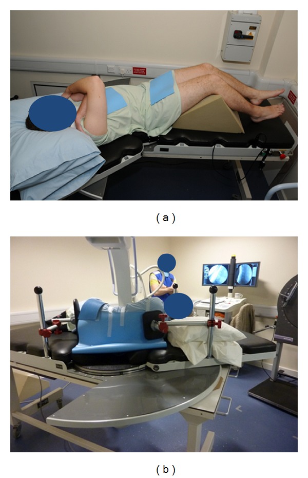
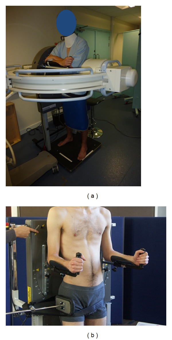
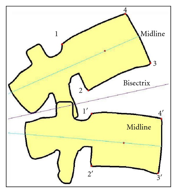
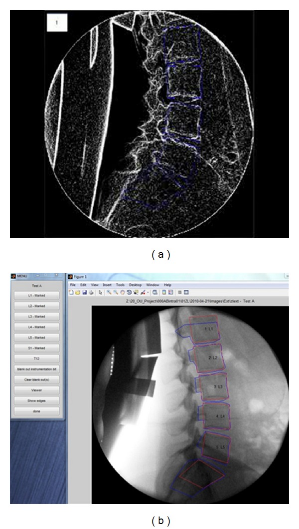
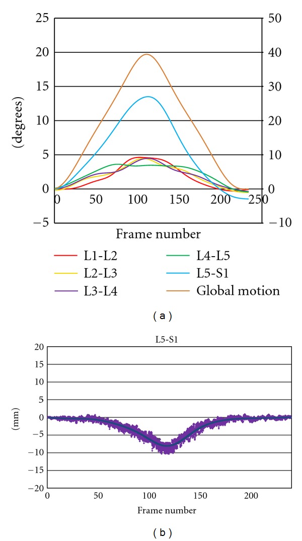
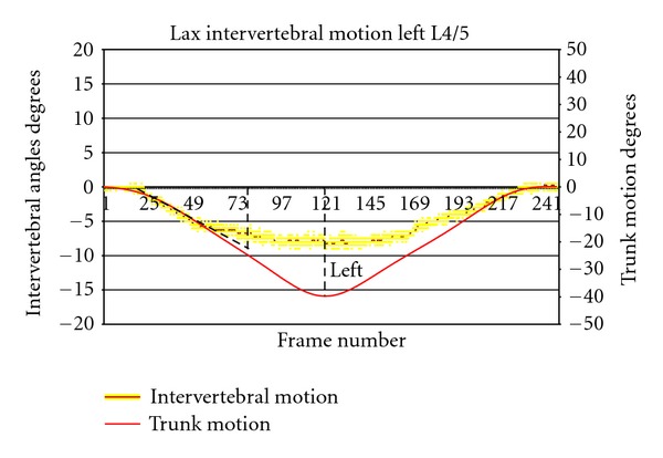
Quantitative fluoroscopy (QF) is an emerging technology for measuring intervertebral motion patterns to investigate problem back pain and degenerative disc disease. This International Forum was a networking event of three research groups (UK, US, Hong Kong), over three days in San Francisco in August 2009. Its aim was to reach a consensus on how best to record, analyse, and communicate QF information for research and clinical purposes. The Forum recommended that images should be acquired during regular trunk motion that is controlled for velocity and range, in order to minimise externally imposed variability as well as to correlate intervertebral motion with trunk motion. This should be done in both the recumbent passive and weight bearing active patient configurations. The main recommended outputs from QF were the true ranges of intervertebral rotation and translation, neutral zone laxity and the consistency of shape of the motion patterns. The main clinical research priority should initially be to investigate the possibility of mechanical subgroups of patients with chronic, nonspecific low back pain by comparing their intervertebral motion patterns with those of matched healthy controls.
Figures
References
-
- Fick R. Handbuch der Anatomie und Mechank der Gelenke. Siena, Italy: Fischer Verlage; 1904.
-
- Todd TW, Pyle IS. A quantitative study of the vertebral column by direct roentgenologic methods. American Journal of Physical Anthropology. 1928;12:321–338.
-
- Gianturco C. A roentgen alalysis of the motion of the lower lumbar vertebrae in normal individuals and in patients with low back pain. American Journal of Roentgenology. 1944;52(3):261–268.
-
- Hasner E, Schalimtzek M, Snorrason E. Roentgenological examination of the function of the lumbar spine. Acta Radiologica. 1952;37(2):141–149. - PubMed
-
- Miles M, Sullivan WE. Lateral bending at the lumbar and lumbosacral joints. The Anatomical Record. 1961;139(3):387–398.
LinkOut - more resources
Full Text Sources
