. 2012:2012:459806.
doi: 10.1155/2012/459806. Epub 2012 May 16.

Alterations in Lipid Levels of Mitochondrial Membranes Induced by Amyloid-β: A Protective Role of Melatonin

Affiliations

Alterations in Lipid Levels of Mitochondrial Membranes Induced by Amyloid-β: A Protective Role of Melatonin

Sergio A Rosales-Corral et al. Int J Alzheimers Dis. 2012.

Abstract

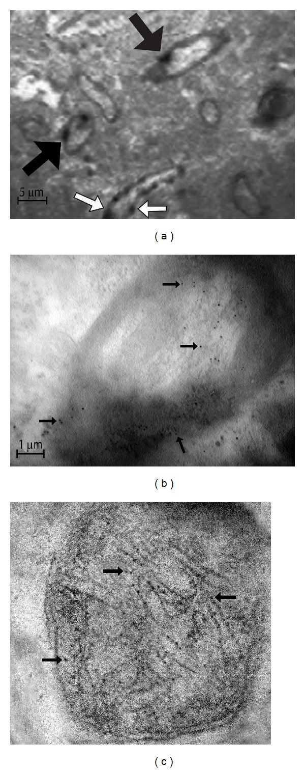
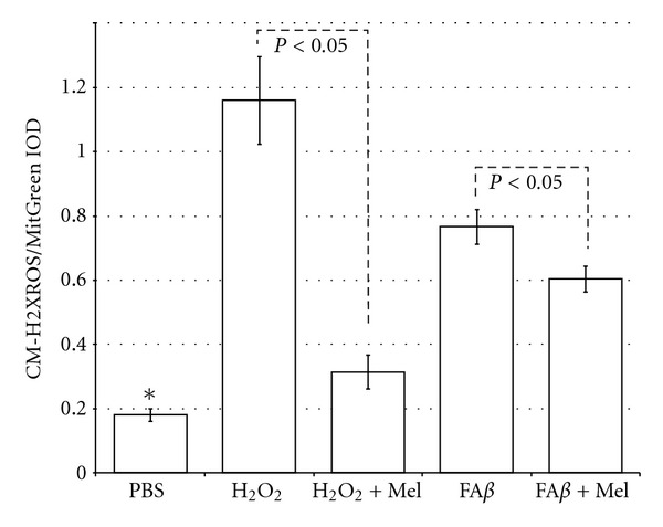
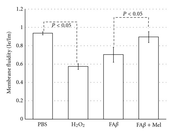
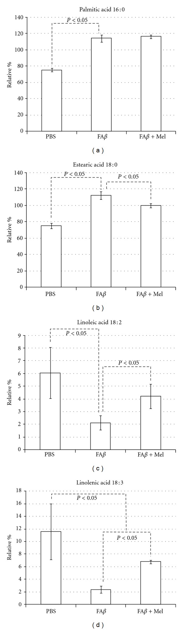
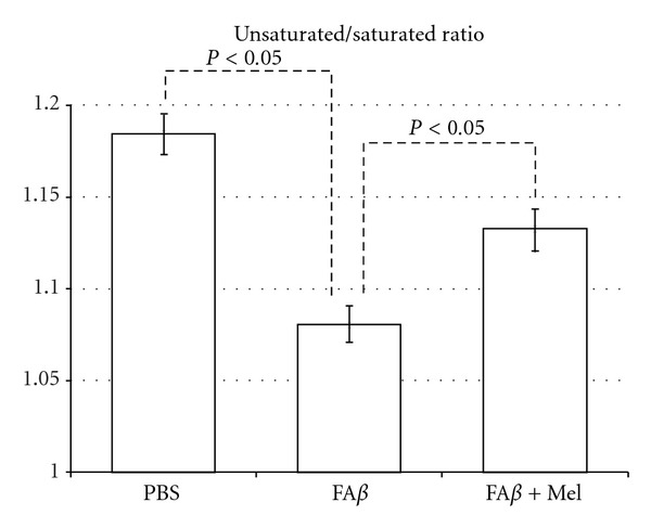
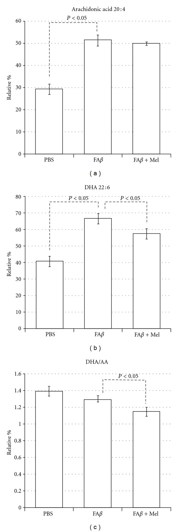
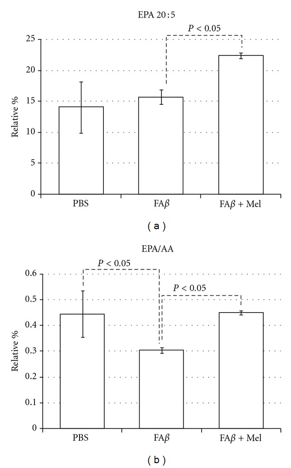
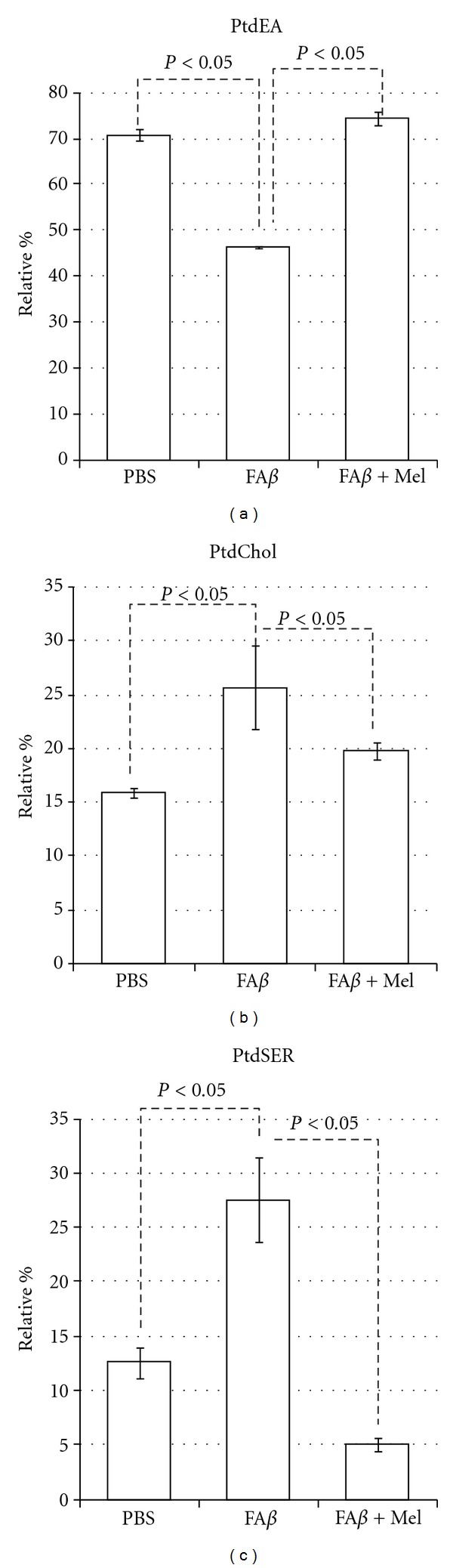
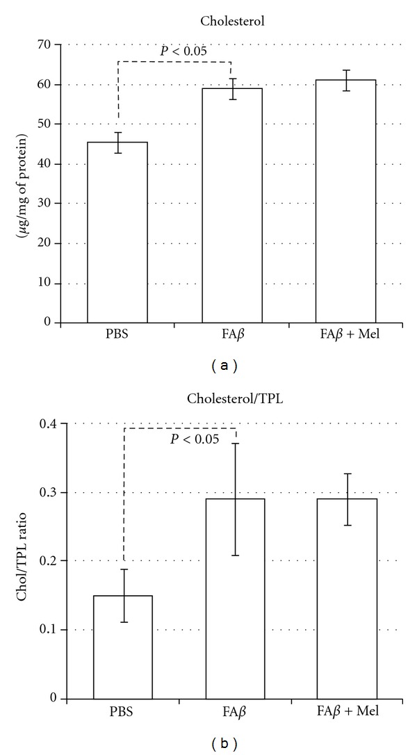
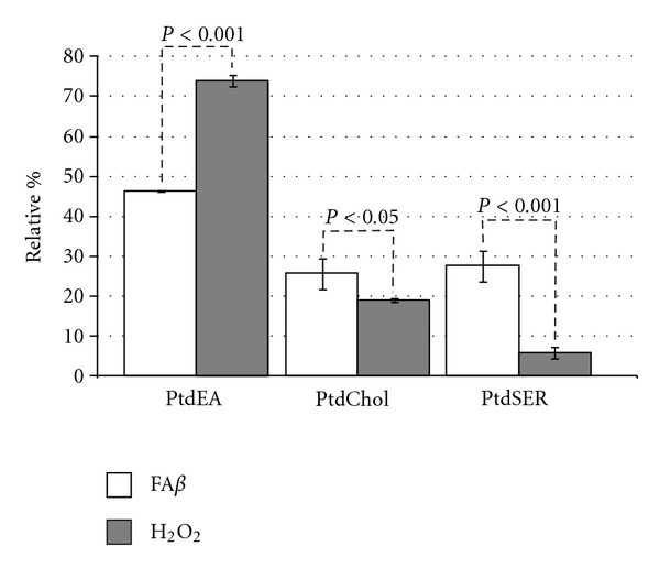
Alzheimer pathogenesis involves mitochondrial dysfunction, which is closely related to amyloid-β (Aβ) generation, abnormal tau phosphorylation, oxidative stress, and apoptosis. Alterations in membranal components, including cholesterol and fatty acids, their characteristics, disposition, and distribution along the membranes, have been studied as evidence of cell membrane alterations in AD brain. The majority of these studies have been focused on the cytoplasmic membrane; meanwhile the mitochondrial membranes have been less explored. In this work, we studied lipids and mitochondrial membranes in vivo, following intracerebral injection of fibrillar amyloid-β (Aβ). The purpose was to determine how Aβ may be responsible for beginning of a vicious cycle where oxidative stress and alterations in cholesterol, lipids and fatty acids, feed back on each other to cause mitochondrial dysfunction. We observed changes in mitochondrial membrane lipids, and fatty acids, following intracerebral injection of fibrillar Aβ in aged Wistar rats. Melatonin, a well-known antioxidant and neuroimmunomodulator indoleamine, reversed some of these alterations and protected mitochondrial membranes from obvious damage. Additionally, melatonin increased the levels of linolenic and n-3 eicosapentaenoic acid, in the same site where amyloid β was injected, favoring an endogenous anti-inflammatory pathway.


Figures

Figure 1
Figure 1
Aβ stain by immunoelectron microscopy. 36 hours after the intracerebral injection of Aβ tissues from the injected area were obtained and subjected to immunohistochemistry by using a primary polyclonal antibody against Aβ. Deposits of Aβ forming deposits in the extracellular space were revealed by conventional light microscopy (data not shown). Aβ immunoreactivity was then revealed with a 6 nm gold label and observed in a transmission electron microscope which allows us to identify (a) deposits of Aβ within myelin axons (black arrows) and in the vasculature (white arrows). (b) Deposits of Aβ (black arrows) penetrate the axon membranes causing demyelination and appear in the axons. Axons look like bulb onions. (c) Aβ appears within the mitochondria finally, where it forms deposits along the cristae (black arrows) and causes intense inflammation, destruction of membranes, and vacuolization (magnification at 27800x).
Figure 2
Figure 2
Compared with PBS-injected brains, those brains injected with Aβ or with H2O2 had a significant increase in free radical levels in mitochondria, according to the CM-H2XROS/MitGreen quotient (*P < 0.05 versus all the other groups). However, by using melatonin a significant decrease in mitochondrial free radicals was observed both in Aβ- and in H2O2-injected brains.
Figure 3
Figure 3
Brains of animals injected with Aβ showed a significant reduction in membrane fluidity as compared with PBS-injected brains, although less obvious than the observed in H2O2-injected brains, used as a positive control, which is in concordance with the degree of the free radicals overproduction, as shown in the previous graphic. Membrane fluidity in animals receiving melatonin was restored at the same level than the PBS group.
Figure 4
Figure 4
Aβ and H2O2 (not shown) had similar and highly significant effects on saturated fatty acids particularly on palmitic and estearic acids whose percentages were increased 39 and 37% correspondingly. Linoleic acid was reduced to a third from the control, while linolenic acid was reduced to less than a quart from the control value, as shown. These important effects of Aβ on specific saturated and unsaturated fatty acids affected the unsaturated/saturated (U/S) balance.
Figure 5
Figure 5
fAβ-injected brains decreased significantly the U/S ratio, as compared with the PBS-injected brains. However, brains of animals taking oral melatonin showed a U/S ratio closer to the control group.
Figure 6
Figure 6
Aβ and H2O2 (not shown) produced important increases in both n6 and n3 PUFA, which reflects the previous described changes in free fatty acids. A similar increase in DHA and AA allowed the DHA/AA ratio to remain stable, when compared with the PBS group. It is obvious that melatonin reduces the DHA/AA ratio, particularly at the expense of a decrease in DHA levels.
Figure 7
Figure 7
EPA was not to significantly responsive to Aβ. However, in the presence of melatonin and contrary to the results with the other major n3 PUFA, DHA, the relative percentage of EPA rose significantly, which impacted the EPA/AA ratio, as shown.
Figure 8
Figure 8
Aβ decreased significantly the PtdEA levels and increased the levels of PtdCHOL and PtdSER, the latter with a 5-fold increment. Results are expressed in relative percentage ± standard error.
Figure 9
Figure 9
Cholesterol content in mitochondrial membranes is significantly increased in fAβ injected brains. The H2O2 control group (data not shown) and the fAβ experimental group, which showed the more important overproduction of free radicals and the lowest membrane fluidity, coincide with the highest cholesterol content. However, in spite of its ability to scavenge free radicals and restore membrane fluidity, melatonin was unable to reduce cholesterol content in mitochondrial membrane. Compared according to their relative values, cholesterol and total phospholipids ratio was significantly altered.
Figure 10
Figure 10
Significant differences between Aβ-injected brains and H2O2-injected brains.

References

    1. De Pietri Tonelli D, Mihailovich M, Di Cesare A, Codazzi F, Grohovaz F, Zacchetti D. Translational regulation of BACE-1 expression in neuronal and non-neuronal cells. Nucleic Acids Research. 2004;32(5):1808–1817. - PMC - PubMed
    1. Maggio JE, Mantyh PW. Brain amyloid—a physicochemical perspective. Brain Pathology. 1996;6(2):147–162. - PubMed
    1. Yatin SM, Varadarajan S, Link CD, Butterfield DA. In vitro and in vivo oxidative stress associated with Alzheimer’s amyloid β-peptide (1–42) Neurobiology of Aging. 1999;20(3):325–342. - PubMed
    1. Rosales-Corral S, Tan DX, Reiter RJ, et al. Orally administered melatonin reduces oxidative stress and proinflammatory cytokines induced by amyloid-β peptide in rat brain: a comparative, in vivo study versus vitamin C and E. Journal of Pineal Research. 2003;35(2):80–84. - PubMed
    1. Lassmann H. Mechanisms of neurodegeneration shared between multiple sclerosis and Alzheimer’s disease. Journal of Neural Transmission. 2011;118(5):747–752. - PubMed

LinkOut - more resources