

Hepatic metastases of primary jejunal carcinoid tumor: A case report with radiological findings
- PMID: 22666712
- PMCID: PMC3364631
Hepatic metastases of primary jejunal carcinoid tumor: A case report with radiological findings
Abstract
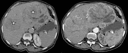
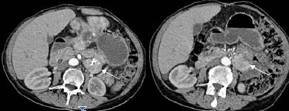
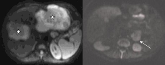
Context: Carcinoid tumors represent a group of well-differentiated tumors originating from the diffuse endocrine system outside the pancreas and thyroid. The overall prevalence of carcinoid tumors in the United States is estimated to be one to two cases per 100,000 persons. Various sites of origin of this neoplasm are appendix - 30-45%, small bowel - 25-35% (duodenum 2%, jejunum 7%, ileum 91%, multiple sites 15-35%), rectum 10-15%, caecum - 5%, and stomach - 0.5%. Liver metastases from jejunal and ileal carcinoids are generally hypervascular.
Case report: Here we report a case of primary jejunal carcinoid tumor in a 66-year-old woman metastasizing to liver with ultrasonography, computed tomography, and diffusion-weighted magnetic resonance imaging (DWI) findings.
Conclusion: Primary jejunal carcinoid tumor is a rare entity. DWI can help in the differential diagnosis of hepatic hypervascular metastatic mass lesions from benign ones, as well as in the diagnosis of carcinoid tumor.
Keywords: Carcinoid; diffusion weighted MRI; jejunum; metastases; small bowel.
Figures
References
-
- Modlin IM, Sandor A. An analysis of 8305 cases of carcinoid tumors. Cancer. 1997;79(4):813–829. - PubMed
-
- Modlin IM, Lye KD, Kidd M. A 5-decade analysis of 13,715 carcinoid tumors. Cancer. 2003;97(4):934–959. - PubMed
-
- Maglinte DDT, Herlinger H. Small bowel neoplasms. In: Herlinger H, Maglinte DDT, Birnbaum BA, editors. Clinical imaging of the small intestine. 2nd ed. New York, NY: Springer; 1999. pp. 377–438.
-
- Ichikawa T, Haradome H, Hachiya J, Nitatori T, Araki T. Diffusion-weighted MR imaging with a single-shot echoplanar sequence: detection and characterization of focal hepatic lesions. AJR Am J Roentgenol. 1998;170(2):397–402. - PubMed
Publication types
LinkOut - more resources
Full Text Sources