Case Reports
. 2009 Nov;1(6):309-13.

Presence of neutrophil extracellular traps and antineutrophil cytoplasmic antibodies associated with vasculitides

Affiliations
Case Reports

Presence of neutrophil extracellular traps and antineutrophil cytoplasmic antibodies associated with vasculitides

Ana Maria Abreu-Velez et al. N Am J Med Sci. 2009 Nov.

Abstract

Context: Anti-neutrophil p-ANCA antibodies are directed against antigens in the peripheral cytoplasm of both neutrophilic granulocytes and monocytes. They are detected in several autoimmune disorders and are particularly associated with systemic vasculitis.

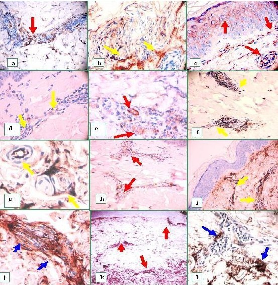
Case report: We report a case of a 54-year-old female presenting with a pruritic rash, including purpura and diffuse erythema. A biopsy with hematoxylin and eosin (H & E) analysis, direct immunofluorescence (DIF), immunohistochemistry (IHC) and enzyme-linked immunosorbent assays for ANCAs were performed. The H & E staining demonstrated leukocytoclastic vasculitis, with focal vascular fibrinoid necrosis. The DIF revealed evidence of vasculitis, the presence of p-ANCAs and neutrophil extracellular traps (NETs). The IHC displayed autoreactivity to myeloperoxidase within the vessels. The IHC aided in ruling out any intrinsic autofluorescence of the vessels.

Conclusions: By observing the deposition of neutrophil extracellular traps and myeloperoxidase in inflamed skin vessels, biopsy analysis may alert physicians for rapid therapeutic intervention in patients presenting with possible vasculitides.

Keywords: IgD; Vasculitis; myeloperoxidase; neutrophil extracellular traps (NETs); p-ANCA; skin.


Figures

Fig. 1
Fig. 1
Presence of neutrophil extracellular traps and antineutrophil cytoplasmic antibodies associated with vasculitides using DIF and IHC. a through f, and i, DIF. a. Area of vasculitis, showing deposits of FITC-conjugated anti-human fibrinogen within dermal vessels (red arrows). b. Intra-cytoplasmic deposits of FITC-conjugated antihuman C1q within epidermal cells (red arrow). c. Positive p-ANCAs (yellow arrow) using FITC-conjugated anti-human IgG. The nuclei are counterstained in red with TO-PRO-3 dye (blue arrow); vessel autoreactivity is displayed in green utilizing the FITC-conjugated anti-human IgG (white arrow). d. Positive p-ANCAs (yellow arrows) in the dermis utilizing FITC-conjugated anti-human IgG antibodies. The nuclei are counterstained in red with TO-PRO-3 (blue arrows). e, Same as d, but at higher magnification, and without nuclear counterstaining. f. Shows a detail of the p-ANCA antigen(s) in the epidermis and the NETs (yellow arrows). g IHC staining with collagen IV shows strong reactivity to the BMZ, as well as around the superficial dermal vessels (brown stain) (aqua arrows). h. H & E staining demonstrating leukocytoclastic vasculitis with fibrinoid necrosis (blue arrow). .i, DIF, and g, deposits of nuclear dust (yellow) on some superficial vessels utilizing FITC-conjugated anti-human IgA antibodies (yellow arrows). The blue arrows show the nuclei counterstained with TO-PRO-3. j An IHC stain using anti-CD34 on the vascular area in i, and showing strong, fragmented endothelial expression of this molecule (aqua arrows). k, IHC stain utilizing myeloperoxidase (red arrows). l. IHC CD31 antibody shows fragmented endothelial cells (red arrows).
Fig. 2
Fig. 2
Real immunoreactivity versus intrinsic, non-pathologic background immunoreactivity in the dermal blood vessels using IHC. Our results confirmed our previous DIF findings. In panel a, note fragmented positivity for CD 34 (red arrow). In b, autoreactivity to intermediate vessels using C1q (yellow arrows). c. Again using C1q, 1) an intracytoplasmic epidermal stratum spinosum reactivity is noted [(also shown with the DIF], as well as 2) a reactivity to the superficial plexus dermal vessels, and 3) to the intermediate dermal vessels (red arrows), (also IHC colocalized with both anti-CD31/PECAM and CD34 antibodies). d. Interestingly, some of these vessels also demonstrated positive findings utilizing anti-human IgD (yellow arrows) e, (red arrows). In f, Dermal perivascular reactivity was also noted utilizing anti-IgE (yellow arrows) and utilizing IgA (data not shown). g. Autoreactivity to the vessels utilizing anti-IgM (yellow arrows), as well as in h, to IgG (red arrows). In (i) note positive autoreactivity to the basal BMZ area, and the superficial vascular plexus (yellow arrows), and, in j, to intermediate dermal vessels (blue arrows). Finally, in k and l, we note compartmentalization or “tight cuffing” of the observed in the inflammatory areas utilizing anti-human fibrinogen (red arrows), and anti-human albumin (blue arrows), respectively.

References

    1. Radice A, Sinico RA. Antineutrophil cytoplasmic antibodies (ANCA) Autoimmunity. 2005;38:93–103. - PubMed
    1. Carlson JA, Ng BT, Chen KR. Cutaneous vasculitis update: diagnostic criteria, classification, epidemiology, etiology, pathogenesis, evaluation and prognosis. Am J Dermatopathol. 2005;27:504–528. - PubMed
    1. Reumaux D, Duthilleul P, Roos D. Pathogenesis of diseases associated with antineutrophil cytoplasm autoantibodies. Hum Immunol. 2004;65:1–12. - PubMed
    1. Kain R, Matsui K, Exner M, Binder S, Schaffner G, Sommer EM, Kerjaschki D. A novel class of autoantigens of anti-neutrophil cytoplasmic antibodies in necrotizing and crescentic glomerulonephritis: the lysosomal membrane glycoprotein h-lamp-2 in neutrophil granulocytes and a related membrane protein in glomerular endothelial cells. J Exp Med. 1995;181:585–597. - PMC - PubMed
    1. Henry JB. 19th ed. Vol. 1556. Philadelphia: W.B. Saunders; 1996. Clinical Diagnosis and Management by Laboratory Methods; pp. 992–993.

Publication types

LinkOut - more resources