. 2012 Jun 27;134(25):10664-9.
doi: 10.1021/ja303737a. Epub 2012 Jun 15.

In vivo fluorescence imaging in the second near-infrared window with long circulating carbon nanotubes capable of ultrahigh tumor uptake

Affiliations

In vivo fluorescence imaging in the second near-infrared window with long circulating carbon nanotubes capable of ultrahigh tumor uptake

Joshua T Robinson et al. J Am Chem Soc. .

Abstract

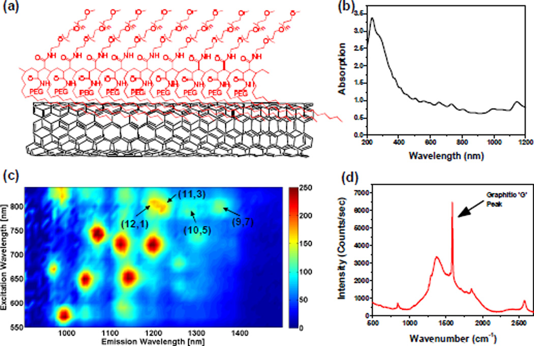
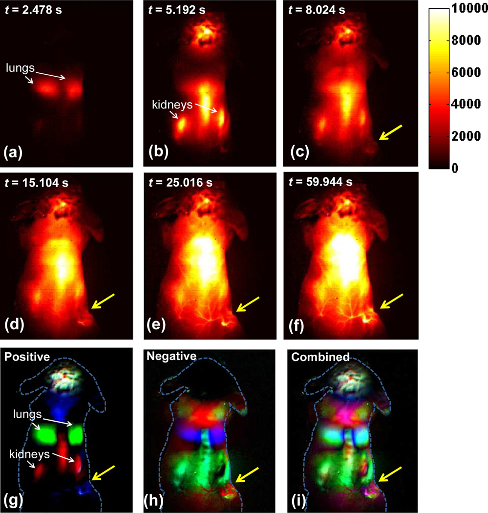
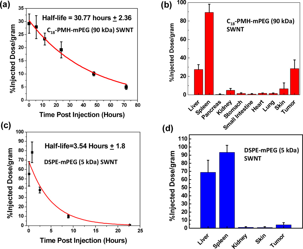
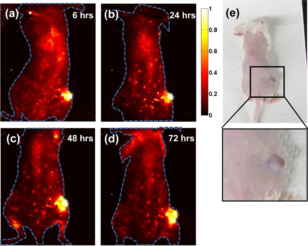
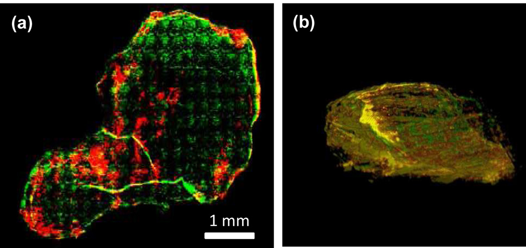
Cancer imaging requires selective high accumulation of contrast agents in the tumor region and correspondingly low uptake in healthy tissues. Here, by making use of a novel synthetic polymer to solubilize single-walled carbon nanotubes (SWNTs), we prepared a well-functionalized SWNT formulation with long blood circulation (half-life of ∼30 h) in vivo to achieve ultrahigh accumulation of ∼30% injected dose (ID)/g in 4T1 murine breast tumors in Balb/c mice. Functionalization dependent blood circulation and tumor uptake were investigated through comparisons with phospholipid-PEG solubilized SWNTs. For the first time, we performed video-rate imaging of tumors based on the intrinsic fluorescence of SWNTs in the second near-infrared (NIR-II, 1.1-1.4 μm) window. We carried out dynamic contrast imaging through principal component analysis (PCA) to immediately pinpoint the tumor within ∼20 s after injection. Imaging over time revealed increasing tumor contrast up to 72 h after injection, allowing for its unambiguous identification. The 3D reconstruction of the SWNTs distribution based on their stable photoluminescence inside the tumor revealed a high degree of colocalization of SWNTs and blood vessels, suggesting enhanced permeability and retention (EPR) effect as the main cause of high passive tumor uptake of the nanotubes.


Figures

Figure 1
Figure 1
Characterization of C18-PMH-mPEG (90 kDa) coated SWNTs. (a) A schematic of the water soluble SWNT conjugate (b) A UV-Vis-NIR absorption spectrum of the suspension (c) A 2D photo-excitation and emission (PLE) map of a C18-PMH-mPEG (90 kDa) coated SWNTs, with different chirality SWNTs showing up as bright spots. (d) A Raman scattering spectrum of the SWNT suspension excited by a 785 nm laser. The graphitic band (G band) is indicated by an arrow at ~1600 cm−1.
Figure 2
Figure 2
Time course NIR-II fluorescence images and dynamic contrast-enhanced images based on PCA analysis. (a–f) NIR-II fluorescence images of a 4T1 tumor bearing mouse after injection of a 200 µL solution containing 0.35 mg/mL SWNTs. (g) Positive pixels from PCA, showing lungs, kidneys and major vessels in the tumor (h) Negative pixels from PCA, showing the body of the tumor. (i) Overlaid image showing the absolute value of both positive and negative pixels, from which both the vessels in the tumor and the tumor outline can be seen. Yellow arrows in images highlight the tumor.
Figure 3
Figure 3
a) Concentration of SWNTs (black symbols) in blood vs. time measured for an injected 90 kDa C18-PMH-mPEG SWNT solution (see Methods section). A first order exponential decay model was used for fitting (red curve). b) Biodistribution of the 90 kDa C18-PMH-mPEG SWNTs in various organs measured by collecting organs from the mice (n=4) 100 hours after injection (see Methods). c) Circulation and d) biodistribution of DSPE-mPEG (5 kDa) coated SWNT in vivo (n=3).
Figure 4
Figure 4
NIR-II imaging of xenograft 4T1 tumor with high uptake of SWNTs (a–d) Time course NIR-II fluorescence images of the same mouse injected with C18-PMH-mPEG (90 kDa) coated SWNTs, showing increasing tumor contrast due to the accumulation of nanotubes inside the tumor. (e) A digital camera image of the same mouse as shown in 4a–d, with noticeable darkening of the tumor due to the high tumor uptake of SWNTs.
Figure 5
Figure 5
Ex vivo imaging of 4T1 murine tumor slices with high SWNT uptake. (a) A tumor slice from a tumor showing the location of SWNTs (coded in red) and Cy5-labeled anti-mouse CD31 (coded in green), and their colocalization (yellow). (b) A reconstructed 3D snapshot image taken from Supplementary Movie 2 with the same color coding as in (a).

References

    1. Wagner V, Dullaart A, Bock AK, Zweck A. Nature Biotechnol. 2006;24:1211–1217. - PubMed
    1. Jain RK, Stylianopoulos T. Nature Rev. Clin. Oncol. 2010;7:653–664. - PMC - PubMed
    1. Stroh M, Zimmer JP, Duda DG, Levchenko TS, Cohen KS, Brown EB, Scadden DT, Torchilin VP, Bawendi MG, Fukumura D, Jain RK. Nature Med. 2005;11:678–682. - PMC - PubMed
    1. Allen PM, Liu W, Chauhan VP, Lee J, Ting AY, Fukumura D, Jain RK, Bawendi MG. J. Am. Chem. Soc. 2009;132:470–471. - PMC - PubMed
    1. Duncan R. Nature Rev. Cancer. 2006;6:688–701. - PubMed

Publication types