. 2012 Aug 17;7(8):1367-76.
doi: 10.1021/cb300216f. Epub 2012 Jun 5.

Bipartite tetracysteine display reveals allosteric control of ligand-specific EGFR activation

Affiliations

Bipartite tetracysteine display reveals allosteric control of ligand-specific EGFR activation

Rebecca A Scheck et al. ACS Chem Biol. .

Abstract

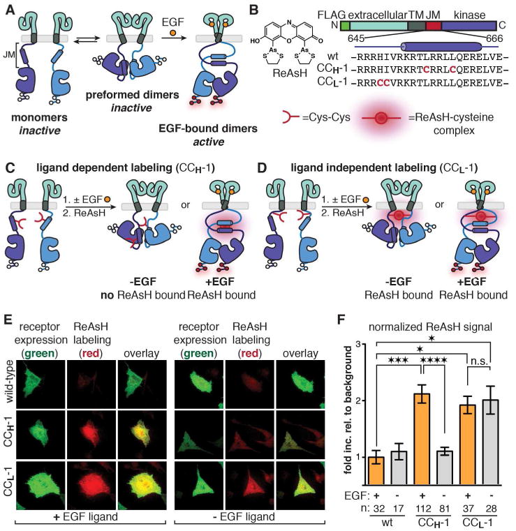
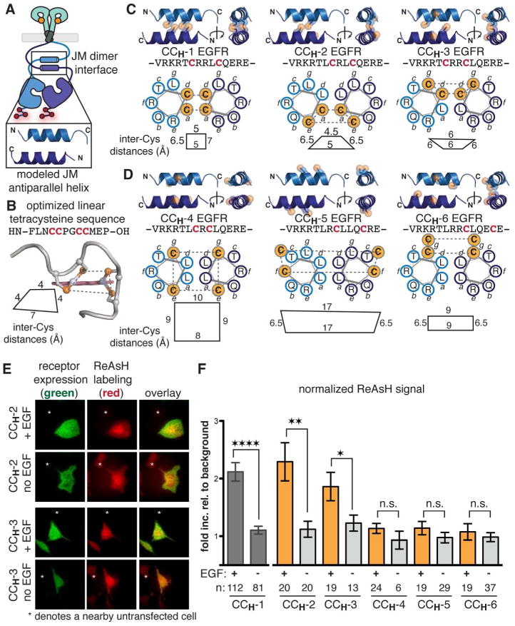
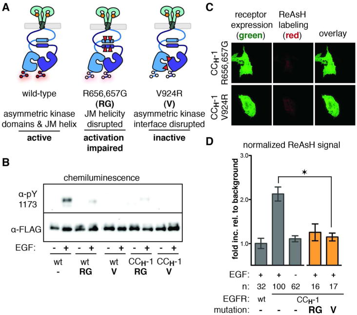
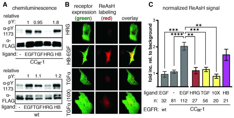
Aberrant activation of the epidermal growth factor receptor (EGFR), a prototypic receptor tyrosine kinase, is critical to the biology of many common cancers. The molecular events that define how EGFR transmits an extracellular ligand binding event through the membrane are not understood. Here we use a chemical tool, bipartite tetracysteine display, to report on ligand-specific conformational changes that link ligand binding and kinase activation for full-length EGFR on the mammalian cell surface. We discover that EGF binding is communicated to the cytosol through formation of an antiparallel coiled coil within the intracellular juxtamembrane (JM) domain. This conformational transition is functionally coupled to receptor activation by EGF. In contrast, TGFα binding is communicated to the cytosol through formation of a discrete, alternative helical interface. These findings suggest that the JM region can differentially decode extracellular signals and transmit them to the cell interior. Our results provide new insight into how EGFR communicates ligand-specific information across the membrane.


Figures

Figure 1
Figure 1. Monitoring EGFR Dimerization and Activation Using Bipartite Tetracysteine Display
(a) Cartoon depicting the current model for EGFR activation. White circles represent sites where tyrosine becomes phosphorylated (red circles) when the receptor is activated. (b) Chemical structure of ReAsH along with the domain structure of EGFR and the identities of two JM Cys-Cys constructs prepared. (c) Cartoon demonstrating EGF-dependent ReAsH labeling of CCH-1 EGFR. (d) Cartoon depicting ligand independent labeling of CCL-1 EGFR. (e) Representative TIRFM images for monitoring the ReAsH labeling of wild-type, CCH-1, and CCL-1 EGFR in the presence (left) and absence (right) of EGF. (f) Quantification of TIRFM results as a fold increase relative to background that is normalized for receptor expression levels. n is the number of cells quantified. Error bars represent the standard error. *p<0.05, *** p<0.001, **** p<0.0001, one-way ANOVA with Bonferroni post-analysis accounting for multiple comparisons. ReAsH labeling of CCH-1 EGFR is dependent on the presence of EGF, whereas ReAsH labeling of CCL-1 can occur regardless of ligand. These data support the conclusion that an interhelical JM interaction is present upon EGF binding.
Figure 2
Figure 2. A JM antiparallel helical dimer is present when EGFR is stimulated with EGF
(a) Cartoon representation of the proposed antiparallel coiled coil and the modeled coordinates for this interaction. (b) Structure of an optimized linear tetracysteine peptide in complex with ReAsH. Inter-cysteine distances are measured from the sulfur atoms. (c) Bipartite Cys-Cys EGFR variants expected to be labeled with ReAsH if the proposed antiparallel coiled coil is formed (see also Supporting Figure S5). Inter-cysteine distances are measured from the sulfur atoms. (d) Bipartite Cys-Cys EGFR variants not expected to be labeled with ReAsH due to an unfavorable binding site geometry when the coiled coil is present. Inter-cysteine distances are measured from the sulfur atoms. (e) Representative TIRFM images of cells expressing CCH-2 and CCH-3 EGFR treated with ReAsH in the presence and absence of EGF. TIRFM images for CCH-4, CCH-5, and CCH-6 can be found in Supporting Figure S4. (f) Quantification of TIRFM results as a fold increase relative to background that is normalized for receptor expression levels. n is the number of cells quantified. Error bars represent the standard error. * p< 0.05, ** p< 0.01, and **** p< 0.0001, t-test analysis. The ReAsH labeling results for a series of Cys-Cys variants provide further evidence that the JM interacts through an antiparallel coiled coil when EGFR is stimulated with EGF. Additional analysis (Supporting Figure S5) rules out parallel association in the two most likely registers.
Figure 3
Figure 3. ReAsH Labeling of the JM Antiparallel Helices is Linked to a Global Active Conformation
(a) Cartoon depicting the relative positions of the activation-impairing EGFR mutations R656,657G and V924R. (b) Western blots confirm that these mutants are defective in tyrosine autophosphorylation in the context of wild-type and CCH-1 EGFR. (c) Representative TIRFM images of ReAsH-treated cells expressing CCH-1 EGFR variants containing the R656,657G or V924R mutations. (d) Quantification of TIRFM results as a fold increase relative to background that is normalized for receptor expression levels. Error bars represent the standard error. * represents p<0.05 based on ANOVA with Bonferroni post-test. The ability of CCH-1 to bind ReAsH is dependent on the presence of JM helices (R656,657G) and the global active conformation of kinase domains (V924R). The absence of ReAsH labeling in these variants provides a structural link between the receptor activation and formation of an antiparallel JM coiled coil.
Figure 4
Figure 4. TGFα activates EGFR through an alternative orientation of JM helices
(a) Western blot analysis of wild-type and CCH-1 EGFR stimulated with different growth factor ligands. (b) Representative TIRFM images for ReAsH labeling of CCH-1 EGFR in the presence of HRG, HB-EGF or TGFα. (c) Quantification of TIRFM results as a fold increase relative to background normalized for receptor expression levels. n is the number of cells quantified. Error bars represent the standard error. ** p<0.01, *** p<0.001, **** p<0.0001, one-way ANOVA with Bonferroni post-analysis accounting for multiple comparisons. The activation of EGFR by TGFα does not involve the JM antiparallel coiled coil that was observed for activation by EGF.
Figure 5
Figure 5. TGFα activates EGFR through a distinct orientation of JM helices
(a) Western blot analysis of wt, R656,657G, and V924R EGFR stimulated with EGF or TGFα. See also Figure 3A. (b) Western blot analysis of CCH-5 and CCH-6 EGFR stimulated with EGF or TGFα. (c) Representative TIRFM images of cells expressing CCH-5 and CCH-6 EGFR that were labeled with ReAsH in the presence or absence of TGFα. See also Supporting Figure S4. (d) Quantification of TIRFM results as a fold increase relative to background that is normalized for receptor expression levels. n is the number of cells quantified. Error bars represent the standard error. ** p<0.01, *** p<0.001, **** p<0.0001, one-way ANOVA with Bonferroni post-analysis accounting for multiple comparisons. TGFα leads to a structural transition in the JM helices, allowing for CCH-5 and CCH-6 to be labeled with ReAsH. These findings suggest that activation of EGFR by TGFα occurs through a JM helical orientation that is distinct from the antiparallel coiled coil determined for activation by EGF.
Figure 6
Figure 6. The Relative Orientation of Domain IV is Different when TGFα is Bound Instead of EGF
(a) Four views of the crystal structure of the EGFR extracellular domain (aqua) bound to EGF (orange). (b) Four views of the homology model of the EGFR extracellular domain (gray) bound to TGFα (yellow). Red residues represent a steric clash observed in domain IV of the homology model. (c) Overlay of the two structures aligned. Comparison reveals a difference in the orientation of domain IV depending on the ligand identity. This analysis is consistent with a model in which differential signaling by TGFα may propagate through a change in the relative orientation of the TM, which results in structural differences in the JM domain.

References

    1. Lemmon MA, Schlessinger J. Cell signaling by receptor tyrosine kinases. Cell. 2010;141(7):1117–1134. - PMC - PubMed
    1. Avraham R, Yarden Y. Feedback regulation of EGFR signalling: decision making by early and delayed loops. Nature Reviews Molecular Cell Biology. 2011;12(2):104–117. - PubMed
    1. Lemmon MA. Ligand-induced ErbB receptor dimerization. Experimental Cell Research. 2009;315(4):638–648. - PMC - PubMed
    1. Tao RH, Maruyama IN. All EGF(ErbB) receptors have preformed homo- and heterodimeric structures in living cells. Journal Of Cell Science. 2008;121(19):3207–3217. - PubMed
    1. Zhang X, Gureasko J, Shen K, Cole PA, Kuriyan J. An allosteric mechanism for activation of the kinase domain of epidermal growth factor receptor. Cell. 2006;125(6):1137–1149. - PubMed

Publication types