

doi: 10.2144/0000113878.
Trehalose-enhanced isolation of neuronal sub-types from adult mouse brain
Affiliations
- PMID: 22668417
- PMCID: PMC3696583
- DOI: 10.2144/0000113878
Item in Clipboard
Trehalose-enhanced isolation of neuronal sub-types from adult mouse brain
Biotechniques.
2012 Jun.
Abstract
Efficient isolation of specific, intact, living neurons from the adult brain is problematic due to the complex nature of the extracellular matrix consolidating the neuronal network. Here, we present significant improvements to the protocol for isolation of pure populations of neurons from mature postnatal mouse brain using fluorescence activated cell sorting (FACS). The 10-fold increase in cell yield enables cell-specific transcriptome analysis by protocols such as nanoCAGE and RNA seq.
Conflict of interest statement
The authors declare no competing interests.
Figures
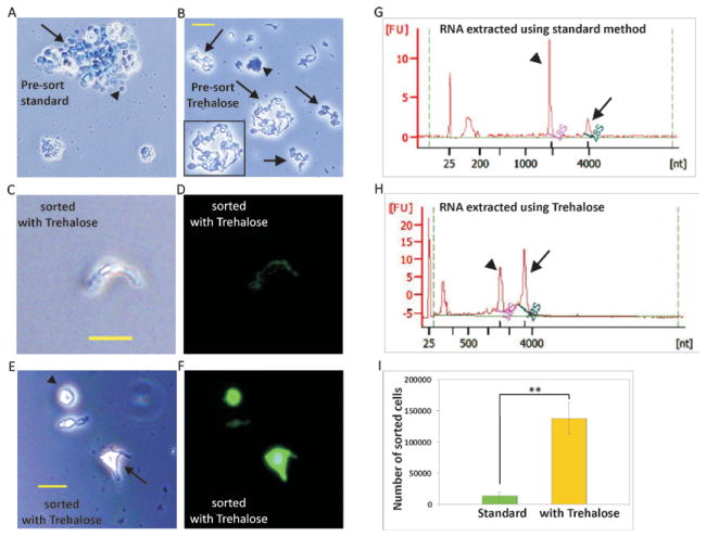
Images taken before sorting (A–B) for standard protocol (A) and optimized protocol with Trehalose (B). Note the dead cells in undissociated tissue (arrow) as well as dead single cells without projections (arrowhead) in the standard protocol. Dissociated intact live neurons (arrows and inset) as well as dead dissociated neurons (arrowhead) are seen with the optimized protocol. (C–F) Images taken after sorting for optimized protocol. Note the intact neuron (C–D) and cells with small (arrow) or no projections (arrowhead) (E–F) in the after sort images from optimized protocol. Scale bar 20μm. Bioanalyzer profiles of RNA extracted from sorted pyramidal neurons isolated using the standard protocol (G) and optimized protocol with Trehalose (H). Note the size of the 28S peak (arrow) is smaller than the 18S peak (arrowhead) in the RNA extracted using the standard method, suggesting RNA degradation with the standard protocol. (I) Chart depicting the yields of fluorescent pyramidal cells using the standard method (n = 2) and our optimized method (n = 6), (Cell numbers shown in Tables 1 and 2). Cell numbers are increased 10-fold when trehalose is used. ** P < 0.001; Student’s t-test, error bars represent s.d.
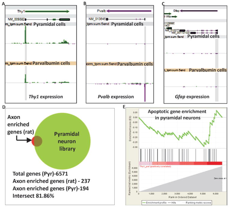
(A–C) Expression levels of Thy1, Pvalb and Gfap in libraries prepared using our optimized protocol. Each snapshot shows Refseq transcripts in the top section and tags per million (tpm) in libraries in the middle and bottom sections. Fixed expression scale of 1000 is shown for all panels. Pyramidal cell libraries (n = 12) and parvalbumin cell libraries (n = 6) as labeled. Tags at the transcript start site (TSS) are shaded in gray. Thy1 expression is found in both pyramidal and parvalbumin cells. Pvalb expression is seen in Parvalbumin libraries alone and Gfap expression is not seen in either of the libraries. (D) Graphical representation of the intersection between axon-enriched mRNAs cataloged in rat (237 genes) (38) and our adult mouse pyramidal neuron nanoCAGE libraries (6571 genes), showing the presence of 194 axons enriched genes in our pyramidal libraries. (E) GSEA plot depicting the enrichment score of genes up-regulated in programmed cell death, Enrichment score (ES) −0.29 and Normalized Enrichment Score (NES) −0.96. The analysis reveals that our pyramidal neuron libraries are not enriched in genes up-regulated during programmed cell death.
References
-
- Dougherty JD, Geschwind DH. Progress in realizing the promise of microarrays in systems neurobiology. Neuron. 2005;45:183–185. - PubMed
-
- Saxena A, Carninci P. Whole transcriptome analysis: what are we still missing? Wiley Interdiscip Rev Syst Biol Med. 2011;3:527–543. - PubMed
-
- Feng G, et al. Imaging neuronal subsets in transgenic mice expressing multiple spectral variants of GFP. Neuron. 2000;28:41–51. - PubMed
Publication types
MeSH terms
Substances
Grants and funding
LinkOut - more resources
Full Text Sources
Other Literature Sources