. 2012 Jun 5;106(12):1989-96.
doi: 10.1038/bjc.2012.196.

FADD protein release mirrors the development and aggressiveness of human non-small cell lung cancer

Affiliations

FADD protein release mirrors the development and aggressiveness of human non-small cell lung cancer

Y Cimino et al. Br J Cancer. .

Abstract

Background: The need to unfold the underlying mechanisms of lung cancer aggressiveness, the deadliest cancer in the world, is of prime importance. Because Fas-associated death domain protein (FADD) is the key adaptor molecule transmitting the apoptotic signal delivered by death receptors, we studied the presence and correlation of intra- and extracellular FADD protein with development and aggressiveness of non-small cell lung cancer (NSCLC).

Methods: Fifty NSCLC patients were enrolled in this prospective study. Intracellular FADD was detected in patients' tissue by immunohistochemistry. Tumours and distant non-tumoural lung biopsies were cultured through trans-well membrane in order to analyse extracellular FADD. Correlation between different clinical/histological parameters with level/localisation of FADD protein has been investigated.

Results: Fas-associated death domain protein could be specifically downregulated in tumoural cells and FADD loss correlated with the presence of extracellular FADD. Indeed, human NSCLC released FADD protein, and tumoural samples released significantly more FADD than non-tumoural (NT) tissue (P=0.000003). The release of FADD by both tumoural and NT tissue increased significantly with the cancer stage, and was correlated with both early and late steps of the metastasis process.

Conclusion: The release of FADD by human NSCLC could be a new marker of poor prognosis as it correlates positively with both tumour progression and aggressiveness.


Conflict of interest statement

The authors declare no conflict of interest.

Figures

Figure 1
Figure 1
Loss of FADD in ex vivo NSCLC can result from FADD release. (AC) Fas-associated death domain protein immunohistochemistry analysis of NT (A) and T tissues (B and C). T0, neg; T1, + T2, ++ T3, +++. A, alveolar; B, bronchial; EC, epithelial cells; Cy, cytoplasm; Nu, nucleus; T, tumour. (D) According to the patients' cancer stage, in vitro T biopsies' FADD release (extracellular FADD) determined by ELISA and ex vivo tumour cells' intracellular FADD expression determined by IHC were inversely correlated (n=24 patients, all from the Hôtel-Dieu). Data represented mean ±s.e.m. Intracellular FADD expression concerns tumour cells only (not normal epithelial cells). Stage I, II, III: n=5, 6, and 13, respectively.
Figure 2
Figure 2
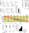
Fas-associated death domain protein is released by human NSCLC. The amount of FADD released by T (black) and by distant NT (white) biopsies in vitro was determined by ELISA. (A) Fas-associated death domain protein release by different samples (diamond) of the same biopsy is shown for seven patients. Bars represent the mean value for each patient. Histogram represents the mean s.d. of FADD release for T and NT patients. (B) Fas-associated death domain protein release by T and NT biopsies from 50 NSCLC patients. When several samples of a same biopsy were tested, we used the samples' mean value. Each diamond represents a patient. Bars represent the mean value for the entire cohort. P values were determined using the Wilcoxon signed-rank test. Range for T and NT biopsies were from 5.3 to 339.7 and 1.2 to 122.5 ng mg−1 PT, respectively. (C) Spearman’s correlation. (D and E) Fas-associated death domain protein release depends on the NSCLC histological type. Levels of released FADD determined in Figure 2B were reanalysed after the 50 NSCLC patients were classified according to their histological type. Each diamond represents a patient biopsy (T in black, NT in white). Bars represent the mean value (D). Histograms represent the mean released FADD value ±s.e.m. P values were determined using the Mann–Whitney test (E).
Figure 3
Figure 3
Fas-associated death domain protein release is correlated with cancer development. (AC) Fas-associated death domain protein release is correlated with patients' cancer stage. Levels of released FADD determined in Figure 2B were reanalysed after the 50 NSCLC patients were classified according to their cancer stage. Each diamond represents a patient biopsy (T in black, NT in white). Bars represent the mean value. P values were determined using the Wilcoxon signed-rank test (A). Histograms represent the mean released FADD value ±s.e.m. P values were determined using the Mann–Whitney test (B). (D) Patient 89 is a 54-year-old smoking (60 pack per year) man. Surgical resection of brain metastasis was performed in 2007, followed by 2 years of radiotherapy and chemotherapy. Fas-associated death domain protein release by remitted tumour (dark grey) and adjacent parenchyma (light grey) biopsies following 1-h in vitro culture was determined by ELISA, and was compared with the mean level of FADD released by the 50 NSCLC T (black) and NT (white) biopsies. Abbreviation: NS=not significant.
Figure 4
Figure 4
Fas-associated death domain protein release is correlated with cancer aggressiveness. (AC) Fas-associated death domain protein release by human NSCLC is positively correlated with the presence of vascular emboli and the lymph node invasion. (A) For 26 patients of the cohort, the level of released FADD is correlated with the presence (+, n=22) or absence (−, n=4) of vascular emboli. For other patients, histopathological report gave no information on the presence or absence of emboli. (B and C) Levels of released FADD determined in Figure 2B were reanalysed after the 50 NSCLC patients were classified according to their lymph node invasion status. Each diamond represents a patient biopsy (T in black, NT in white). Bars represent the mean value. n represents the number of patients; one patient with N3 NSCLC is not represented. P values were determined using the Wilcoxon signed-rank test (B). Histograms represent the mean released FADD value ±s.e.m. of biopsies from N0 (n=23) and N+ (n=26) patients (C). P values were determined using the Mann–Whitney test (A, C). Abbreviation: NS=not significant.

References

    1. Alifano M, Cusumano G, Strano S, Magdeleinat P, Bobbio A, Giraud F, Lebeau B, Regnard JF (2009) Lobectomy with pulmonary artery resection: morbidity, mortality, and long-term survival. J Thorac Cardiovasc Surg 137(6): 1400–1405 - PubMed
    1. Balachandran S, Thomas E, Barber GN (2004) A FADD-dependent innate immune mechanism in mammalian cells. Nature 432(7015): 401–405 - PubMed
    1. Beisner DR, Chu IH, Arechiga AF, Hedrick SM, Walsh CM (2003) The requirements for Fas-associated death domain signaling in mature T cell activation and survival. J Immunol 171(1): 247–256 - PubMed
    1. Bell BD, Leverrier S, Weist BM, Newton RH, Arechiga AF, Luhrs KA, Morrissette NS, Walsh CM (2008) FADD and caspase-8 control the outcome of autophagic signaling in proliferating T cells. Proc Natl Acad Sci USA 105(43): 16677–16682 - PMC - PubMed
    1. Bhojani MS, Chen G, Ross BD, Beer DG, Rehemtulla A (2005) Nuclear localized phosphorylated FADD induces cell proliferation and is associated with aggressive lung cancer. Cell Cycle 4(11): 1478–1481 - PubMed

Publication types

Substances