. 2012 Aug 1;303(3):E410-21.
doi: 10.1152/ajpendo.00039.2012. Epub 2012 Jun 5.

JAK/STAT3 pathway inhibition blocks skeletal muscle wasting downstream of IL-6 and in experimental cancer cachexia

Affiliations

JAK/STAT3 pathway inhibition blocks skeletal muscle wasting downstream of IL-6 and in experimental cancer cachexia

Andrea Bonetto et al. Am J Physiol Endocrinol Metab. .

Abstract

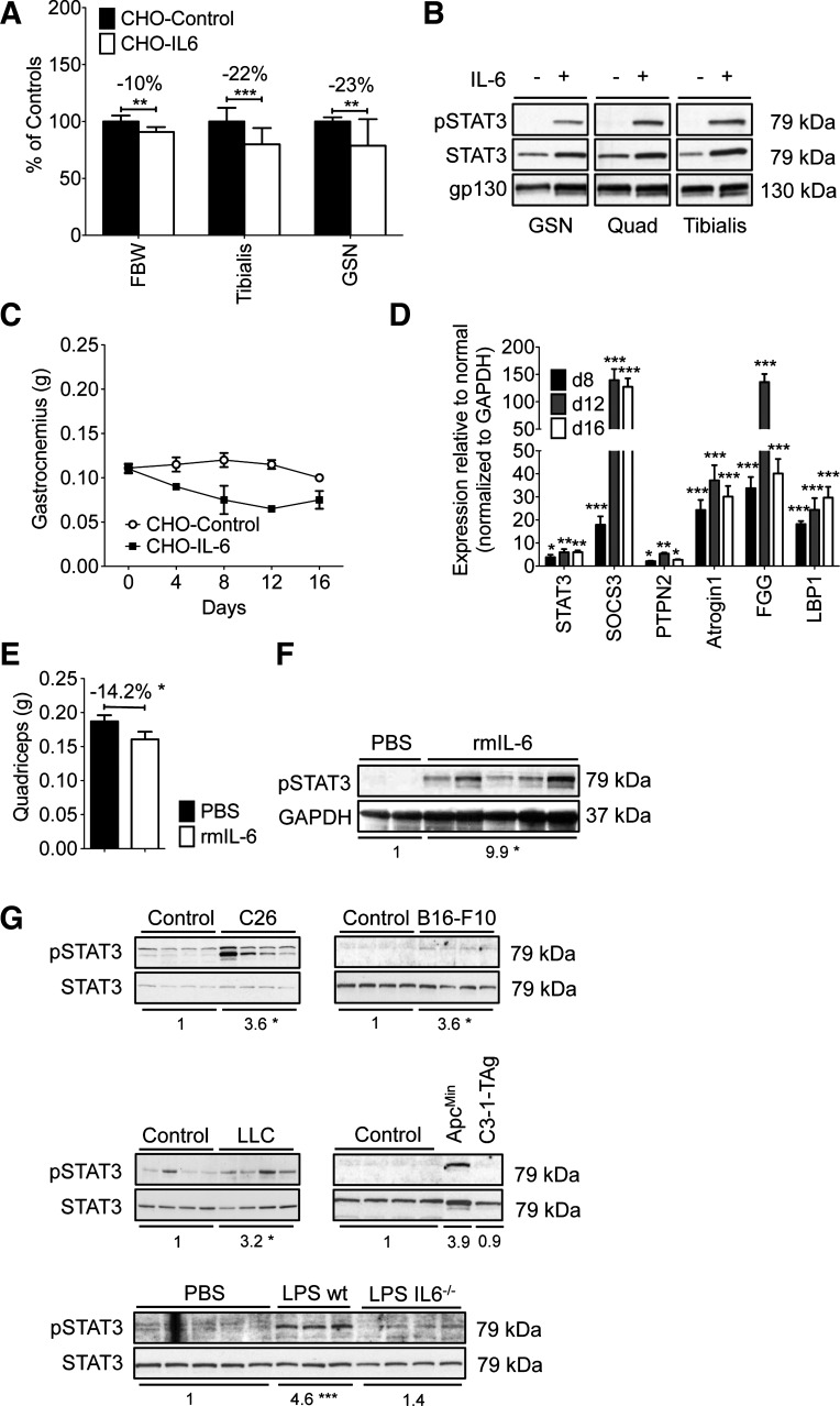
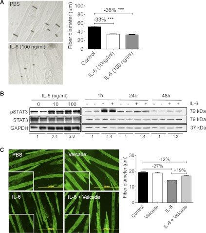
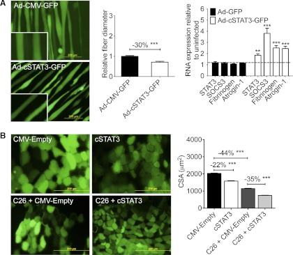
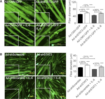
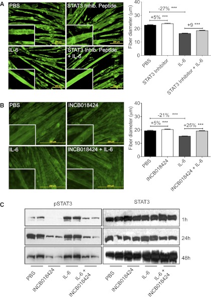
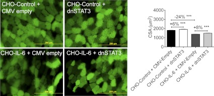
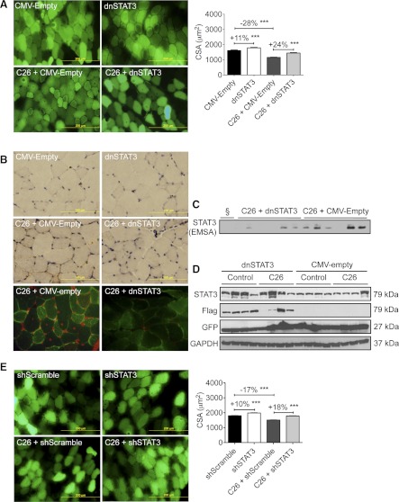
Cachexia, the metabolic dysregulation leading to sustained loss of muscle and adipose tissue, is a devastating complication of cancer and other chronic diseases. Interleukin-6 and related cytokines are associated with muscle wasting in clinical and experimental cachexia, although the mechanisms by which they might induce muscle wasting are unknown. One pathway activated strongly by IL-6 family ligands is the JAK/STAT3 pathway, the function of which has not been evaluated in regulation of skeletal muscle mass. Recently, we showed that skeletal muscle STAT3 phosphorylation, nuclear localization, and target gene expression are activated in C26 cancer cachexia, a model with high IL-6 family ligands. Here, we report that STAT3 activation is a common feature of muscle wasting, activated in muscle by IL-6 in vivo and in vitro and by different types of cancer and sterile sepsis. Moreover, STAT3 activation proved both necessary and sufficient for muscle wasting. In C(2)C(12) myotubes and in mouse muscle, mutant constitutively activated STAT3-induced muscle fiber atrophy and exacerbated wasting in cachexia. Conversely, inhibiting STAT3 pharmacologically with JAK or STAT3 inhibitors or genetically with dominant negative STAT3 and short hairpin STAT3 reduced muscle atrophy downstream of IL-6 or cancer. These results indicate that STAT3 is a primary mediator of muscle wasting in cancer cachexia and other conditions of high IL-6 family signaling. Thus STAT3 could represent a novel therapeutic target for the preservation of skeletal muscle in cachexia.


Figures

Fig. 1.
Fig. 1.
IL-6-induced and tumor-induced muscle wasting were associated with STAT3 activation in vivo and in vitro. A: Chinese hamster ovary (CHO)/IL-6 cells injected in athymic nude mice caused loss of body weight [final body weight (FBW)] and muscle weight, including tibialis anterior and gastrocnemius vs. CHO/control mice (n = 8/group, euthanized on day 12). Data are representative of >10 experiments. B: Western blotting analysis of muscle from mice in A reveals increased Y705-STAT3 in the gastrocnemius (GSN), quadriceps (Quad), and tibialis anterior of mice treated with CHO/IL-6 cells (+IL-6) vs. CHO/controls (−IL-6). Blot is representative of 4 independently assayed samples from each group on day 8, but virtually identical results were observed on days 4 and 12 (not shown). C: muscle wasting was progressive over time with CHO/IL-6 cells vs. CHO/controls. Gastronemius is shown (n = 4–6/point). D: quantitative real-time RT-PCR (qPCR) shows elevated expression of STAT3 target genes and atrogin-1 in quadriceps from mice with cachexia and CHO/IL-6 from days 8, 12, and 16 normalized to CHO/control samples (n = 3/condition, sampled in triplicate). E and F: recombinant IL-6 administered by osmotic pumps (1 μg/h for 7 days) induced a marked reduction in quadriceps weight (n = 4–6/group; E) and increased pY705-STAT3 by Western blotting analysis of mice euthanized after 7 days of treatment (F). This experiment is 1 of 2. Fold change vs. control group (PBS) for the pSTAT3/GAPDH ratio is shown under the blots (*P < 0.05). G: quadriceps pY705-STAT3 was increased in experimental models of cancer cachexia when weight loss in the tumor-bearing group was equal to 10% of starting body weight for C26, B16-F10, and Lewis lung carcinoma (LLC)-tumor bearing mice and ApcMin mice or after 7 days of chronic LPS administration by osmotic pump. Note that IL-6-null (IL6−/−) mice showed less pSTAT3 with LPS pumps. Fold change vs. control group for the pSTAT3/STAT3 ratio is reported under each of the blots. *P < 0.05; **P < 0.01; ***P < 0.001.
Fig. 2.
Fig. 2.
IL-6 induced loss of C2C12 myofiber mass and activated STAT3 in a proteasome-dependent manner. A: IL-6 treatment resulted in loss of C2C12 myofiber diameter after 48 h. Black bars show representative myotube diameter (n = 158–200 fibers/condition from 3 independent wells). Data are representative of >8 experiments. ***P < 0.001. B: in C2C12 myotubes, increased pSTAT3 was observed after 1 h of treatment with 10 or 100 ng/ml IL-6 (left) and after 1, 24, and 48 h of 100 ng/ml IL-6. Fold change vs. respective control group for the pSTAT3/STAT3 ratio is reported under each set of blots. Data are representative of >5 trials. C: Velcade/bortezomib (1 nM) cotreatment for 48 h prevented IL-6-induced muscle atrophy (n = 170–210 fibers/well from 3 independent wells for each condition).
Fig. 3.
Fig. 3.
STAT3 was sufficient to induce muscle fiber atrophy both in vitro and in vivo. A: constitutively active STAT3 [Ad-cSTAT3-green fluorescent protein (GFP)] resulted in decreased C2C12 myofiber diameter 48 h after infection (n = 150–200 fibers/condition from 3 independent wells) and increased transcription of known STAT3 target genes as well as atrogin-1 (n = 3 wells/group in triplicate). B: cytomegalovirus (CMV)-cSTAT3 transfection reduced cross-sectional area (CSA) in the tibialis anterior muscle of non-tumor-bearing and C26-bearing CD2F1 mice. Mice were transfected on the same day as PBS, or tumor cells were injected and then euthanized 12 days later. Only positively transfected fibers (i.e., green fibers coexpressing pCMV-GFP) were measured (n = 650–1,900 fibers/condition; n = 8 tumor-bearing and non-tumor-bearing mice). Both experiments have been performed >3 times. **P < 0.01; ***P < 0.001.
Fig. 4.
Fig. 4.
STAT3 activity was necessary for muscle atrophy. A: infection of adenovirus expressing dominant negative STAT3 (Ad-dnSTAT3-GFP) resulted in increased fiber diameter in control and IL-6-treated C2C12 myotubes (n = 200–300 fibers from 3 independent wells/condition). B: Ad-shSTAT3-GFP also increased baseline myofiber size and inhibited IL-6-dependent fiber atrophy (n = 200–300 fibers from 3 independent wells/condition). Data represent several independent experiments in which adenovirus was applied for 24 h and then washed out, and IL-6 was applied for 48 h and cells were fixed and measured. ***P < 0.001.
Fig. 5.
Fig. 5.
Pharmacological inhibition of JAK/STAT3 prevented IL-6-dependent myofiber atrophy. A: STAT3 inhibitor peptide, 50 μM, induced modest myofiber hypertrophy and prevented IL-6-induced fiber atrophy after 48 h of cotreatment (n = 200–300 fibers from 3 independent wells/condition). B: the JAK inhibitor INCB018424 (400 nM) induced modest hypertrophy in control myotubes and completely blocked IL-6-mediated myofiber atrophy after 48 h of cotreatment (n = 200–300 fibers from 3 independent wells/condition). Data represent 3 separate experiments. C: Western blotting analysis of whole C2C12 culture extracts show markedly reduced pSTAT3 with INCB018424 treatment at 1, 24, and 48 h. A and B are representative of at least 2 experiments each in which inhibitor and IL-6 were added at the same time and cells were incubated for 48 h and then fixed and measured. ***P < 0.001.
Fig. 6.
Fig. 6.
STAT3 inhibition prevented IL-6-dependent muscle wasting in mice. Expression of dnSTAT3 through CMV-dnSTAT3 transfection of tibialis anterior muscle on the day of CHO injection resulted in basal hypertrophy in CHO/control mice and reduced muscle wasting in CHO/IL-6 mice vs. the CMV-empty vector transfected controls 9 days later (n = 450–1,200 fibers from n = 8 mice/condition, repeated twice). ***P < 0.001.
Fig. 7.
Fig. 7.
STAT3 inhibition using dnSTAT3 or short hairpin (sh)STAT3 prevented C26 tumor-induced muscle atrophy in mice. Mice were transfected on the day of tumor inoculation and euthanized 12 days later. A: CMV-dnSTAT3 transfection of tibialis anterior muscle resulted in basal hypertrophy in non-tumor-bearing mice and reduced muscle wasting in C26 tumor-bearing mice vs. CMV-empty vector transfected controls (n = 250–500 fibers from n = 8 mice/condition, repeated twice). B: immunohistochemistry (brown staining; top) and immunofluorescence (bottom) for pY705-STAT3 in tibialis anterior muscles electroporated with CMV-dnSTAT3. Note pSTAT3 nuclear localization is reduced in the presence of dnSTAT3 (brown staining in immunohistochemistry, red staining in immunofluorescence analyses). C: CMV-dnSTAT3 transfection decreased STAT3 DNA-binding activity in nuclear extracts derived from transfected tibialis muscle in mice with C26 cachexia, as shown by the electrophoretic mobility shift assay (EMSA; n = 6/condition). §Unlabeled probe control. D: Western blotting analysis of total lysate from tibialis of control (non-tumor-bearing) and C26 tumor-bearing mice transfected with CMV-dnSTAT3 or empty vector shows expression of the Flag-taged-dnSTAT3 and GFP, with GAPDH as loading control (n = 4). E: CMV-shSTAT3 increased muscle CSA and prevented C26-induced fiber wasting in the tibialis muscle vs. CMV-shScramble (n = 1,650–2,750 fibers from n = 8 mice/condition, repeated twice). ***P < 0.001.

References

    1. Akiyama Y, Kajimura N, Matsuzaki J, Kikuchi Y, Imai N, Tanigawa M, Yamaguchi K. In vivo effect of recombinant human leukemia inhibitory factor in primates. Jpn J Cancer Res 88: 578–583, 1997 - PMC - PubMed
    1. Alvarez B, Quinn LS, Busquets S, Quiles MT, Lopez-Soriano FJ, Argiles JM. Tumor necrosis factor-alpha exerts interleukin-6-dependent and -independent effects on cultured skeletal muscle cells. Biochim Biophys Acta 1542: 66–72, 2002 - PubMed
    1. Argiles JM, Busquets S, Lopez-Soriano FJ. Cytokines in the pathogenesis of cancer cachexia. Curr Opin Clin Nutr Metab Care 6: 401–406, 2003 - PubMed
    1. Argilés JM, Busquets S, Toledo M, López-Soriano FJ. The role of cytokines in cancer cachexia. Curr Opin Support Palliat Care 3: 263–268, 2009 - PubMed
    1. Baltgalvis KA, Berger FG, Pena MM, Davis JM, Muga SJ, Carson JA. Interleukin-6 and cachexia in ApcMin/+ mice. Am J Physiol Regul Integr Comp Physiol 294: R393–R401, 2008 - PubMed

Publication types

MeSH terms

LinkOut - more resources