

Review
doi: 10.1007/s10439-012-0599-z.
Epub 2012 Jun 6.
Mathematical simulation of membrane protein clustering for efficient signal transduction
Affiliations
- PMID: 22669501
- PMCID: PMC3822010
- DOI: 10.1007/s10439-012-0599-z
Item in Clipboard
Review
Mathematical simulation of membrane protein clustering for efficient signal transduction
Ann Biomed Eng.
2012 Nov.
Abstract
Initiation and propagation of cell signaling depend on productive interactions among signaling proteins at the plasma membrane. These diffusion-limited interactions can be influenced by features of the membrane that introduce barriers, such as cytoskeletal corrals, or microdomains that transiently confine both transmembrane receptors and membrane-tethered peripheral proteins. Membrane topographical features can lead to clustering of receptors and other membrane components, even under very dynamic conditions. This review considers the experimental and mathematical evidence that protein clustering impacts cell signaling in complex ways. Simulation approaches used to consider these stochastic processes are discussed.
Figures
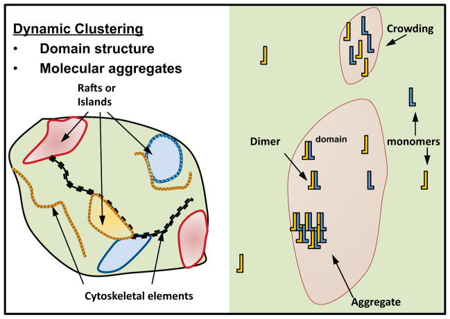
Left: Cartoon representation of features that can subcompartmentalize the plasma membrane, including rafts or islands and the cortical cytoskeletal network. These features are highly dynamic, permitting rapid exchange by diffusion. Right: Representation of the distribution of receptors (yellow, blue symbols) in and out of domains (pink shapes) formed by these features. Arrows point to various states, including monomers, dimers and aggregates. Receptors that are transiently trapped in domains are locally crowded (arrow, top right) and appear as clusters by imaging technologies. This molecular crowding can be more pronounced upon ligand stimulation, due in part to formation of dimers and larger aggregates with decreased diffusive mobility. This review considers the experimental and computational evidence that molecular crowding influences receptor dimerization/aggregation and recruitment of signaling proteins.
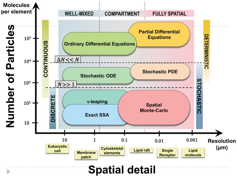
A possible quantitative guide is the size of the largest element that can be treated as spatially homogeneous (horizontal axis) and the typical number of molecules of one species in the element (vertical axis). The suggested spatial resolution is determined by the size of the biological elements of interest and current computational capabilities. Spatially resolved models are resource-intensive, and are therefore generally applied to small subsystems. Cell-level models of large signaling networks are typically well mixed; spatial Monte Carlo studies rarely scale beyond a few hundred nanometers. A promising approach for multi-scale applications is a combination of compartment-based models at the large scales and fully spatial simulations focused on a few important processes within small structural elements of the membrane. Temporal fluctuations arise largely from the discrete and stochastic nature of the underlying molecular processes. The relative magnitude of temporal fluctuations (ΔN) decreases as the number of particles increases. The discrete nature of the particle number can thus be ignored when N is significantly greater than 1. That is, deviations from the expected average behavior can be neglected when the expected magnitude of the fluctuations is small compared to N.
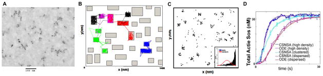
(A). Experimental evidence for EGFR clustering in absence of ligand. Electron micrograph of gold particle-labeled EGF receptors in resting A341 cells (~2 million EGFR/cell), reveals a non-random distribution and provides evidence for receptor co-confinement. (B). Spatial domain used in lattice-free Monte Carlo simulation.[28] The spatial domain simulated by the off-lattice Monte Carlo procedure was a square of area 2 μm2, representative of a small region in the plasma membrane. This region was modified to include many islands or preferred domains (the green rectangles within the membrane patch), to simulate the receptor-trapping micordomains seen in (A). Movement of receptors into and out of the simulated microdomains over a time period of 30 s is indicated by the thin colored tracings. Receptor trapping in the microdomains was reproduced mathematically by stipulating that receptors had a greater probability of entering these regions than of leaving them. (C) Simulation predictions of receptor clustering in absence of ligand. The predicted particle positions after 30 s of simulation time are indicated by the black dots. The Hopkins statistical test (inset) was used to test the randomness of receptor distribution. The right shift of the distribution (compared to the random or normal distribution shown in red) towards unity confirms the clustered nature of the receptors. The predicted receptor distribution compares well with the experimental observation in (A). (D) Simulations using a Coupled Spatial/Nonspatial Stochastic Algorithm (CSNSA) support the conclusion that EGFR clustering promotes activation of the adaptor SOS. ODE models confirm this conclusion, using a fast diffusion coefficient to override contributions from membrane spatial organization. (From Hsieh et al.[28] and Costa et al.)
References
-
- Bublil EM, Yarden Y. The EGF receptor family: spearheading a merger of signaling and therapeutics. Curr Opin Cell Biol. 2007;19(2):124–34. - PubMed
-
- Keating E, Nohe A, Petersen NO. Studies of distribution, location and dynamic properties of EGFR on the cell surface measured by image correlation spectroscopy. Eur Biophys J. 2008;37(4):469–81. - PubMed
-
- Wennerberg K, Rossman KL, Der CJ. The Ras superfamily at a glance. J Cell Sci. 2005;118(Pt 5):843–6. - PubMed
Publication types
MeSH terms
Substances
Grants and funding
LinkOut - more resources
Full Text Sources
