Review
. 2012 Jun;24(6):2279-303.
doi: 10.1105/tpc.112.096586. Epub 2012 Jun 5.

Plant peroxisomes: biogenesis and function

Affiliations
Review

Plant peroxisomes: biogenesis and function

Jianping Hu et al. Plant Cell. 2012 Jun.

Abstract

Peroxisomes are eukaryotic organelles that are highly dynamic both in morphology and metabolism. Plant peroxisomes are involved in numerous processes, including primary and secondary metabolism, development, and responses to abiotic and biotic stresses. Considerable progress has been made in the identification of factors involved in peroxisomal biogenesis, revealing mechanisms that are both shared with and diverged from non-plant systems. Furthermore, recent advances have begun to reveal an unexpectedly large plant peroxisomal proteome and have increased our understanding of metabolic pathways in peroxisomes. Coordination of the biosynthesis, import, biochemical activity, and degradation of peroxisomal proteins allows for highly dynamic responses of peroxisomal metabolism to meet the needs of a plant. Knowledge gained from plant peroxisomal research will be instrumental to fully understanding the organelle's dynamic behavior and defining peroxisomal metabolic networks, thus allowing the development of molecular strategies for rational engineering of plant metabolism, biomass production, stress tolerance, and pathogen defense.


Figures

Figure 1.
Figure 1.
Models for the Biogenesis of Peroxisomes in Plants. In the ER vesiculation model (A), all of the protein constituents of the peroxisomal boundary membrane and matrix are considered to be synthesized cotranslationally on the ER and then sequestered into a specialized region of the ER, where an expanding smooth membrane vesicle eventually buds off to yield (de novo) a nascent, functional peroxisome. By contrast, in the growth and division model (B), all PMPs and matrix proteins are synthesized on free polyribosomes in the cytosol and sorted posttranslationally to preexisting and new (daughter) peroxisomes, resulting in their growth. Daughter peroxisomes arise from preexisting peroxisomes by fission, and the ER somehow provides the membrane lipids necessary for peroxisome growth (e.g., via ER-peroxisome contact sites and/or lipid transfer proteins). In the ER semiautonomous model (C), some PMPs (group I PMPs) are posttranslationally inserted either directly into the pER subdomain or first into the general ER and then routed to the pER. The subsequent transport of these group I PMPs (and membrane lipids) from the pER to preexisting and daughter peroxisomes involves the (de novo) formation (via vesiculation or fragmentation) of putative preperoxisomal carriers that travel to, or from, an ERPIC. All matrix proteins and group II PMPs are sorted posttranslationally from the cytosol to daughter peroxisomes and preexisting peroxisomes, and perhaps preperoxisomes at the ERPIC, the former of which arise by fission (as depicted in more detail in Figure 2).
Figure 2.
Figure 2.
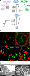
Proteins That Mediate Peroxisome Division in Arabidopsis. (A) A molecular model of peroxisome division in Arabidopsis. Peroxisome elongation is promoted by the PEX11 proteins, among which PEX11b can be transcriptionally activated by light through a phyA-mediated signal transduction pathway. The fission machineries of peroxisomes and mitochondria share at least five components: DRP3A, DRP3B, FIS1A, FIS1B, and PMD1. DRP5B is a common fission factor for peroxisomes and chloroplasts. PMD1 appears to function independently from PEX11 and the FIS1-DRP3 complex by an unknown mechanism. For mitochondrial and chloroplast division, only factors shared with peroxisomes are depicted. (B) Confocal micrographs of leaf mesophyll cells showing peroxisome phenotypes in plants ectopically expressing PEX11a and loss-of-function mutants of DRP3A and DRP5B. WT, the wild type. Bars = 10 µm. (Images reprinted from Orth et al. [2007], Figure 5; Zhang and Hu [2009], Figure 2; Zhang and Hu [2010], Figure 1.) (C) Transmission electron micrographs of leaf mesophyll cells showing the organelle phenotype of plants overexpressing PMD1. Bars = 1 µm. (Images reprinted from Aung and Hu [2011], Figure 4.)
Figure 3.
Figure 3.
Schematic Diagram of Matrix Protein Import into Peroxisomes. Cytosolic PEX5 and PEX7 recognize their cargo proteins (square and round shapes) via binding of specific targeting sequences, PTS1 and PTS2, respectively. Cargo-loaded PEX5 associates with the membrane via interactions with PEX13 and PEX14 and probably also via interactions with the lipid phase. PEX7 cannot dock to the membrane on its own and depends on physical interaction with PEX5 for docking. PTS1- and PTS2-bound cargo is released to the matrix, and the receptors are recycled back into the cytosol via a mechanism that probably requires ATP-dependent ubiquitination of PEX5 (represented by a star) by PEX4 and the RING complex comprised of PEX2, PEX10, and PEX12. The ubiquitinated PEX5 is then removed from the membrane via the action of the AAA ATPases PEX6 and PEX1, which are tethered by APEM9. The route that PEX7 takes through the pathway, in particular whether it accompanies PEX5 throughout the import cycle, is unknown.
Figure 4.
Figure 4.
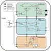
Proteins Acting in Peroxisomal β-Oxidation. (A) Mutants disrupting peroxisomal function frequently have IBA response and Suc-dependent phenotypes. Left, wild-type seedlings grown with applied IBA have shorter primary roots and abundant secondary roots, whereas peroxisome-defective mutants (e.g., pxa1 mutant shown) do not respond to IBA application because of their inability to β-oxidize IBA to IAA. Right, wild-type seedlings germinate and grow normally without an external carbon source, but peroxisome-defective mutants have disruptions in seedling establishment, ranging from failed to delayed development. Bar = 2 mm. (Images reprinted from Zolman et al. [2001], cover photo, and Adham et al. [2005], Figure 8.) (B) Major metabolic pathways in peroxisomes use a core set of enzymes. Fatty acid β-oxidation (center) in developing seeds involves conversion of very-long-chain fatty acids (VLCFA) stored as TAG through long-chain fatty acid (LCFA), medium-chain fatty acid (MCFA), and short-chain fatty acid (SCFA) intermediates. Each round of β-oxidation releases two carbons as acetyl-CoA. IBA (left) and OPDA (right) are metabolized in parallel pathways that use an overlapping but distinct set of enzymes; OPDA is produced from polyunsaturated fatty acids (PUFAs) in a multistep pathway in chloroplasts. *, CTS/PXA1/PED3 may import unmodified substrates or CoA derivatives. For all pathways, substrate activation by acyl-CoA synthetases is shown in purple, the initial oxidation enzymes are in red, the hydration/oxidation intermediate steps (frequently performed by a multifunctional enzyme) are shown in blue, and the thiolysis step is shown in green; if known, specific isozymes catalyzing the reaction are indicated. Peroxisomal acetyl-CoA is a central intermediate in various branches of cellular metabolism, including (1) the conversion to succinate via the glyoxylate cycle, which enters gluconeogenic pathways to produce Glc; (2) the production of malate, necessary for the malate-oxaloacetate shuttle to reoxidize NADH produced by MFP2/AIM1; and (3) the conversion to citrate, which enters the tricarboxylic acid (TCA) cycle.
Figure 5.
Figure 5.
The Central Role of Leaf Peroxisomes in Photorespiration. Photorespiration is compartmentalized among chloroplasts, leaf peroxisomes, mitochondria, and the cytosol. Eleven enzymes are directly involved: Rubisco, phosphoglycolate phosphatases (PGP), GOX, catalase (CAT), Glu:glyoxylate aminotransferase (GGT), Ser:glyoxylate aminotransferase (SGT), Gly decarboxylase (GDC), Ser hydroxymethyl transferase (SHMT), HPR, peroxisomal MDH (pMDH), and GLYK. Four enzymes (i.e., Glu synthase [GS], Glu:oxoglutarate aminotransferase [GOGAT], and mitochondrial/chloroplast malate dehydrogenase [mMDH/cMDH]) are indirectly involved. For the transport of photorespiratory intermediates, different translocators and a porin-like channel have been characterized biochemically (translocators, green; porin-like channel, blue) or cloned (translocator, black). Photorespiratory metabolites are abbreviated as follows: RuBP, ribulose-bisphosphate; 3-PGA, 3-phosphoglycerate; and THF, tetrahydrofolate. (Adapted and reprinted from Reumann and Weber [2006], Figure 1.)

References

    1. Acosta I.F., Farmer E.E. (2010). Jasmonates. The Arabidopsis Book 8: e0129. doi/10.1199/tab.0129
    1. Adham A.R., Zolman B.K., Millius A., Bartel B. (2005). Mutations in Arabidopsis acyl-CoA oxidase genes reveal distinct and overlapping roles in beta-oxidation. Plant J. 41: 859–874 - PubMed
    1. Afitlhile M.M., Fukushige H., Nishimura M., Hildebrand D.F. (2005). A defect in glyoxysomal fatty acid beta-oxidation reduces jasmonic acid accumulation in Arabidopsis. Plant Physiol. Biochem. 43: 603–609 - PubMed
    1. Agrawal G., Joshi S., Subramani S. (2011). Cell-free sorting of peroxisomal membrane proteins from the endoplasmic reticulum. Proc. Natl. Acad. Sci. USA. 108: 9113–9118 - PMC - PubMed
    1. Anand P., Kwak Y., Simha R., Donaldson R.P. (2009). Hydrogen peroxide induced oxidation of peroxisomal malate synthase and catalase. Arch. Biochem. Biophys. 491: 25–31 - PubMed

Publication types

MeSH terms