Case Reports
. 2012;5(4):367-73.
Epub 2012 Apr 16.

Tumor-to-tumor metastasis: pathology and neuroimaging considerations

Affiliations
Case Reports

Tumor-to-tumor metastasis: pathology and neuroimaging considerations

Patricia Moody et al. Int J Clin Exp Pathol. 2012.

Abstract

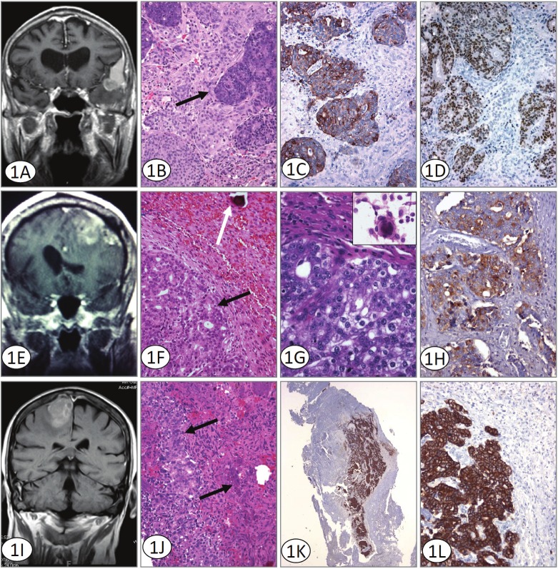
The phenomenon of tumor-to-tumor metastasis has been reported in the literature for over a century. However, it remains fairly uncommon, with fewer than 100 cases being described during that time. Virtually any benign or malignant tumor can be a recipient, but meningiomas have been implicated as the most common intracranial neoplasm to harbor metastasis. The donor neoplasm is most frequently lung or breast carcinoma, while rare cases of metastasis from other primary tumors have been reported. We report here three examples of such rare metastases. This case series reports the first documented instance involving rectal adenocarcinoma. In addition, we report two cases of metastatic prostate adenocarcinoma to a meningioma; to date of which only three cases have been published. The terms "tumor-to-tumor metastasis" and "collision tumor" are addressed, as are details of the pathology. The limitations of standard radiological imaging techniques, such as standard CT and MR, which cannot reliably identify the presence of metastasis within a meningioma are compared with physiology-based neuroimaging methods, such as perfusion MR and MR spectroscopy, which may be more useful in noninvasively differentiating tumor histology.

Keywords: Tumor-to-tumor metastasis; adenocarcinoma; meningioma; neuroimaging; pathology.


Figures

Figure 1
Figure 1
A. Coronal T1 weighted MRI with contrast shows enhancement of the pterional meningioma, with dural tail sign superiorly and metastatic lesion involving the anteromedial part of the tumor. Although radiographically a collision tumor could be considered, histologically islands of metastatic adenocarcinoma surrounded entirely by meningioma are seen. B. H&E stained sections illustrate metastatic colorectal adenocarcinoma (solid arrow) within otherwise typical meningioma. Original magnification 200x. C. Immunohistochemistry for Cytokeratin 20 supports a colorectal origin for the metastatic component defining solid areas and islands of immunoreactive tumor. Cytokeratin 20 immunoreacted sections. Original magnification 200x. D. Immunohistochemistry for Cdx-2 confirms the origin of the tumor and defines solid areas and islands of immunoreactive tumor. Cdx-2 immunoreacted sections. Original magnification 200x. E. MRI revealed a large, 6cm left frontal mass containing blood adjacent to a prominent area of calvarial hyperostosis. Both intra and extra-axial components were identified. The tumor was creating a mass effect with surrounding vasogenic edema. F and G. H&E stained sections illustrate two morphologically distinct areas; an epithelial, glandular component (black arrow) and a second area with a syncytial pattern and a well defined psammoma body (white arrow) is identified. At higher magnification the larger nuclei and prominent nucleoli of the prostatic adenocarcinoma are evident. The inset shows a well-formed meningothelial whorl seen on smear preparation. (Original magnifications 100x and 400x). H. The metastatic adenocarcinoma component of the tumor is prostate specific antibody immunoreactive. Immunohistochemistry for PSAP Original magnification 200x. I. MRI revealed a dural-based, 2.4 x2.6cm lesion located in the area of the right frontal cortex with some vasogenic edema. The lesion displayed some heterogeneity with apparent discrete areas within the tumor which differed from the larger surrounding tumor. J. H&E stained sections illustrate metastatic prostate adenocarcinoma (solid arrow) within meningioma. Original magnification 200x. K and L. The prostate adenocarcinoma component of the tumor demonstrates a discretely positive cytokeratin Cam 5.2 reaction. Immunohistochemistry for Cytokeratin Cam 5.2. Original magnification 100x and 200x.

References

    1. Fried BM. Metastatic Inoculation of a Meningioma by Cancer Cells from a Bronchiogenic Carcinoma. Am J Pathol. 1930;6:47–52. 1. - PMC - PubMed
    1. Petraki C, Vaslamatzis M, Argyrakos T, Petraki K, Strataki M, Alexopoulos C, Sotsiou F. Tumor to tumor metastasis: report of two cases and review of the literature. Int J Surg Pathol. 2003;11:127–135. - PubMed
    1. Lanotte M, Benech F, Panciani PP, Cassoni P, Ducati A. Systemic cancer metastasis in a meningioma: report of two cases and review of the literature. Clin Neurol Neurosurg. 2009;111:87–93. - PubMed
    1. Schmitt HP. Metastases of malignant neoplasms to intracranial tumours: the "tumour-in-a-tumour" phenomenon. Virchows Arch A Pathol Anat Histopathol. 1984;405:155–160. - PubMed
    1. Aghi M, Kiehl TR, Brisman JL. Breast adenocarcinoma metastatic to epidural cervical spine meningioma: case report and review of the literature. J Neurooncol. 2005;75:149–155. - PubMed

Publication types

MeSH terms

Substances

LinkOut - more resources