Review
. 2012 Jun:1257:175-83.
doi: 10.1111/j.1749-6632.2012.06533.x.

Claudins and alveolar epithelial barrier function in the lung

Affiliations
Review

Claudins and alveolar epithelial barrier function in the lung

James A Frank. Ann N Y Acad Sci. 2012 Jun.

Abstract

The alveolar epithelium of the lung constitutes a unique interface with the outside environment. This thin barrier must maintain a surface for gas transfer while being continuously exposed to potentially hazardous environmental stimuli. Small differences in alveolar epithelial barrier properties could therefore have a large impact on disease susceptibility or outcome. Moreover, recent work has focused attention on the alveolar epithelium as central to several lung diseases, including acute lung injury and idiopathic pulmonary fibrosis. Although relatively little is known about the function and regulation of claudin tight junction proteins in the lung, new evidence suggests that environmental stimuli can influence claudin expression and alveolar barrier function in human disease. This review considers recent advances in the understanding of the role of claudins in the breakdown of the alveolar epithelial barrier in disease and in epithelial repair.


Figures

Figure 1
Figure 1
Claudin mRNA expression profiles of primary alveolar epithelial type 1 and type 2 cells. FACS-sorted, freshly-isolated primary rat alveolar epithelial cells predominantly express claudin-3, claudin-4 and claudin-18.1 with transcripts for these claudins accounting for 97% of all claudin transcripts in these cells. In type 1 cells claudin-18.1 is the most abundant transcript, while in type 2 cells claudin-3 is the major transcript. Both cell types express claudin-4. Adapted from reference .
Figure 2
Figure 2
Protein kinase C activation induces claudin-4 expression via a JNK-dependent pathway. Phorbol 12-myristate 13-acetate (PMA), an activator of several PKC isoforms, induces a significant increase in claudin-4 expression in primary in primary human type 2-like distal lung epithelial cells at 4h. This effect was completely inhibited by the protein kinase C inhibitor Gö6850 (not shown). Inhibition of the JNK MAPK pathway with each of three inhibitors (SP600125, AS601245, and JIP-TAT peptide) blocked the PMA-induced increase in claudin 4 expression in a dose-dependent fashion (*P < 0.05 compared with PMA control, data are mean ± SEM, CP = control TAT peptide). Adapted from reference .
Figure 3
Figure 3
Claudin-3 and -4 knock down impairs alveolar fluid clearance and increases lung injury severity in vivo. Clostridium perfringens binding domain peptide (CPE-BD) significantly reduced rates of alveolar fluid clearance by 35–40% in healthy mice. Both basal and beta-adrenergic-stimulated (maximal) rates of alveolar fluid clearance were significantly lower as compared with mice receiving a control peptide (left). Mice given CPE-BD and then exposed to moderate or severe lung injury via escalating tidal volumes on a mechanical ventilator developed more severe pulmonary edema (excess lung water) compared with mice given a control peptide (*P < 0.05 compared with baseline lung water, **P< 0.05 compared with control peptide-treated mice). Despite reduced rates of fluid clearance at baseline, claudin-3 and -4 knock down did not induce pulmonary edema in the absence of an additional injurious stimulus. Adapted from reference .
Figure 4
Figure 4
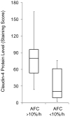
Claudin-4 levels are associated with alveolar fluid clearance rates in human lungs rejected for transplantation. Claudin-4 protein expression levels as assessed by immunostaining were significantly higher in lungs with more preserved alveolar fluid clearance rates. Alveolar fluid clearance was measured in an ex vivo perfused organ system (P < 0.01 for this comparison by Mann-Whitney U). Adapted from reference .

Similar articles

Cited by

References

    1. Tsukita S, Yamazaki Y, Katsuno T, Tamura A. Tight junction-based epithelial microenvironment and cell proliferation. Oncogene. 2008;27:6930–6938. - PubMed
    1. Gorin AB, Stewart PA. Differential permeability of endothelial and epithelial barriers to albumin flux. J Appl Physiol. 1979;47:1315–1324. - PubMed
    1. Vreim CR, Snashall PD, Demling RH, Staub NC. Lung lymph and free interstitial fluid protein composition in sheep with edema. Am J Physiol. 1976;230:1650–1653. - PubMed
    1. Matthay MA, Folkesson HG, Clerici C. Lung epithelial fluid transport and the resolution of pulmonary edema. Physiol Rev. 2002;82:569–600. - PubMed
    1. Matthay MA, Robriquet L, Fang X. Alveolar epithelium: role in lung fluid balance and acute lung injury. Proc Am Thorac Soc. 2005;2:206–213. - PubMed