

The development, distribution and density of the plasma membrane calcium ATPase 2 calcium pump in rat cochlear hair cells
- PMID: 22672315
- PMCID: PMC3412916
- DOI: 10.1111/j.1460-9568.2012.08159.x
The development, distribution and density of the plasma membrane calcium ATPase 2 calcium pump in rat cochlear hair cells
Abstract
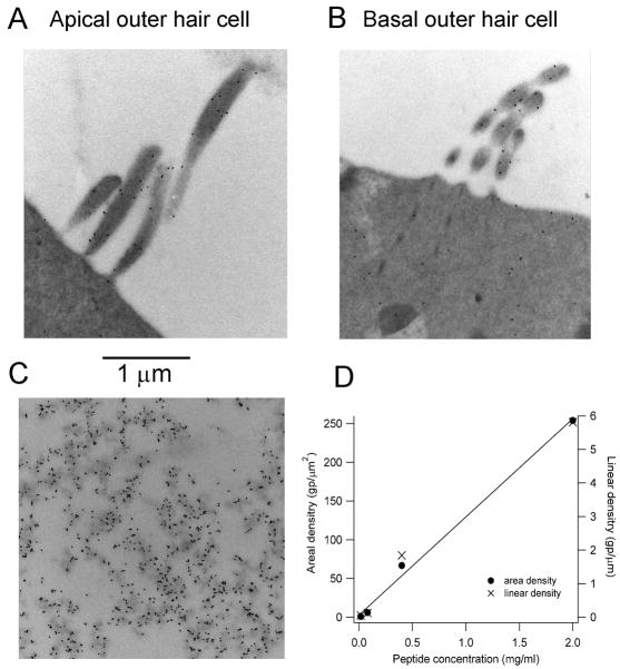
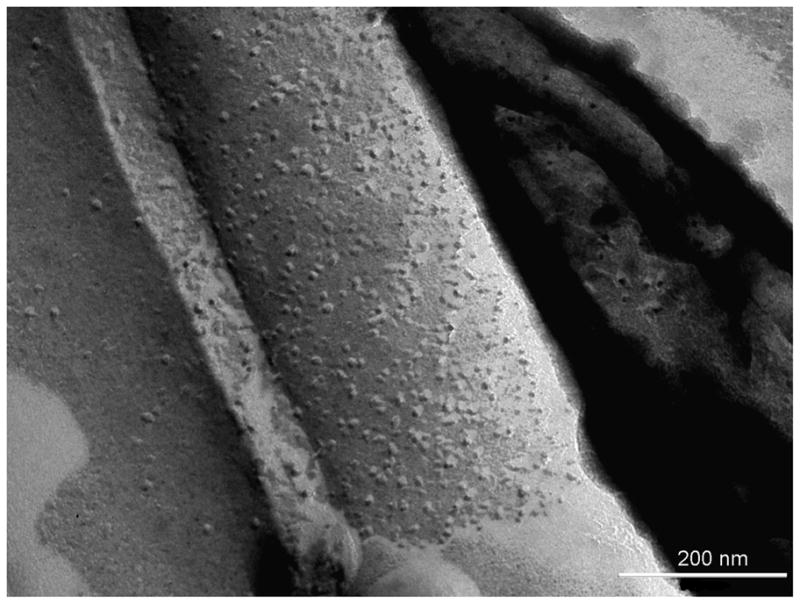
Calcium is tightly regulated in cochlear outer hair cells (OHCs). It enters mainly via mechanotransducer (MT) channels and is extruded by the plasma membrane calcium ATPase (PMCA)2 isoform of the PMCA, mutations in which cause hearing loss. To assess how pump expression matches the demands of Ca(2+) homeostasis, the distribution of PMCA2 at different cochlear locations during development was quantified using immunofluorescence and post-embedding immunogold labeling. The PMCA2 isoform was confined to stereociliary bundles, first appearing at the base of the cochlea around post-natal day (P)0 followed by the middle and then the apex by P3, and was unchanged after P8. The developmental appearance matched the maturation of the MT channels in rat OHCs. High-resolution immunogold labeling in adult rats showed that PMCA2 was distributed along the membranes of all three rows of OHC stereocilia at similar densities and at about a quarter of the density in inner hair cell stereocilia. The difference between OHCs and inner hair cells was similar to the ratio of their MT channel resting open probabilities. Gold particle counts revealed no difference in PMCA2 density between low- and high-frequency OHC bundles despite larger MT currents in high-frequency OHCs. The PMCA2 density in OHC stereocilia was determined in low- and high-frequency regions from calibration of immunogold particle counts as 2200/μm(2) from which an extrusion rate of ∼200 ions/s per pump was inferred. The limited ability of PMCA2 to extrude the Ca(2+) load through MT channels may constitute a major cause of OHC vulnerability and high-frequency hearing loss.
© 2012 The Authors. European Journal of Neuroscience © 2012 Federation of European Neuroscience Societies and Blackwell Publishing Ltd.
Figures


Similar articles
-
Developmental changes in the cochlear hair cell mechanotransducer channel and their regulation by transmembrane channel-like proteins.J Gen Physiol. 2013 Jan;141(1):141-8. doi: 10.1085/jgp.201210913. J Gen Physiol. 2013. PMID: 23277480 Free PMC article.
-
Tonotopy in calcium homeostasis and vulnerability of cochlear hair cells.Hear Res. 2019 May;376:11-21. doi: 10.1016/j.heares.2018.11.002. Epub 2018 Nov 16. Hear Res. 2019. PMID: 30473131 Free PMC article. Review.
-
MET currents and otoacoustic emissions from mice with a detached tectorial membrane indicate the extracellular matrix regulates Ca2+ near stereocilia.J Physiol. 2021 Apr;599(7):2015-2036. doi: 10.1113/JP280905. Epub 2021 Mar 9. J Physiol. 2021. PMID: 33559882 Free PMC article.
-
The ultrastructural distribution of prestin in outer hair cells: a post-embedding immunogold investigation of low-frequency and high-frequency regions of the rat cochlea.Eur J Neurosci. 2010 May;31(9):1595-605. doi: 10.1111/j.1460-9568.2010.07182.x. Eur J Neurosci. 2010. PMID: 20525072 Free PMC article.
-
The plasma membrane calcium pump in the hearing process: physiology and pathology.Sci China Life Sci. 2011 Aug;54(8):686-90. doi: 10.1007/s11427-011-4200-z. Epub 2011 Jul 24. Sci China Life Sci. 2011. PMID: 21786191 Review.
Cited by
-
Mechanotransduction-Dependent Control of Stereocilia Dimensions and Row Identity in Inner Hair Cells.Curr Biol. 2020 Feb 3;30(3):442-454.e7. doi: 10.1016/j.cub.2019.11.076. Epub 2020 Jan 2. Curr Biol. 2020. PMID: 31902726 Free PMC article.
-
Jag1 represses Notch activation in lateral supporting cells and inhibits an outer hair cell fate in the medial cochlea.Development. 2024 Nov 1;151(21):dev202949. doi: 10.1242/dev.202949. Epub 2024 Nov 5. Development. 2024. PMID: 39373109 Free PMC article.
-
Ca2+-pumping by PMCA-neuroplastin complexes operates in the kiloHertz-range.Nat Commun. 2025 Aug 20;16(1):7550. doi: 10.1038/s41467-025-62735-5. Nat Commun. 2025. PMID: 40835599 Free PMC article.
-
Role of Calcium-Sensing Receptor in Mechanotransducer-Channel-Mediated Ca2+ Influx in Hair Cells of Zebrafish Larvae.Front Physiol. 2018 May 30;9:649. doi: 10.3389/fphys.2018.00649. eCollection 2018. Front Physiol. 2018. PMID: 29899708 Free PMC article.
-
Synaptic Contributions to Cochlear Outer Hair Cell Ca2+ Dynamics.J Neurosci. 2021 Aug 11;41(32):6812-6821. doi: 10.1523/JNEUROSCI.3008-20.2021. Epub 2021 Jul 12. J Neurosci. 2021. PMID: 34253627 Free PMC article.
References
-
- Apicella S, Chen S, Bing R, Penniston JT, Llinas R, Hillman DE. Plasmalemmal ATPase calcium pump localizes to inner and outer hair bundles. Neuroscience. 1997;79:1145–1151. - PubMed
Publication types
MeSH terms
Substances
Grants and funding
LinkOut - more resources
Full Text Sources
Miscellaneous
