Enhancement of musculocutaneous nerve reinnervation after vascular endothelial growth factor (VEGF) gene therapy
- PMID: 22672575
- PMCID: PMC3441459
- DOI: 10.1186/1471-2202-13-57
Enhancement of musculocutaneous nerve reinnervation after vascular endothelial growth factor (VEGF) gene therapy
Abstract
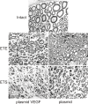
Background: Vascular endothelial growth factor (VEGF) is not only a potent angiogenic factor but it also promotes axonal outgrowth and proliferation of Schwann cells. The aim of the present study was to quantitatively assess reinnervation of musculocutaneous nerve (MCN) stumps using motor and primary sensory neurons after plasmid phVEGF transfection and end-to-end (ETE) or end-to-side (ETS) neurorrhaphy. The distal stump of rat transected MCN, was transfected with plasmid phVEGF, plasmid alone or treated with vehiculum and reinnervated following ETE or ETS neurorrhaphy for 2 months. The number of motor and dorsal root ganglia neurons reinnervating the MCN stump was estimated following their retrograde labeling with Fluoro-Ruby and Fluoro-Emerald. Reinnervation of the MCN stumps was assessed based on density, diameter and myelin sheath thickness of regenerated axons, grooming test and the wet weight index of the biceps brachii muscles.
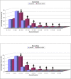
Results: Immunohistochemical detection under the same conditions revealed increased VEGF in the Schwann cells of the MCN stumps transfected with the plasmid phVEGF, as opposed to control stumps transfected with only the plasmid or treated with vehiculum. The MCN stumps transfected with the plasmid phVEGF were reinnervated by moderately higher numbers of motor and sensory neurons after ETE neurorrhaphy compared with control stumps. However, morphometric quality of myelinated axons, grooming test and the wet weight index were significantly better in the MCN plasmid phVEGF transfected stumps. The ETS neurorrhaphy of the MCN plasmid phVEGF transfected stumps in comparison with control stumps resulted in significant elevation of motor and sensory neurons that reinnervated the MCN. Especially noteworthy was the increased numbers of neurons that sent out collateral sprouts into the MCN stumps. Similarly to ETE neurorrhaphy, phVEGF transfection resulted in significantly higher morphometric quality of myelinated axons, behavioral test and the wet weight index of the biceps brachii muscles.
Conclusion: Our results showed that plasmid phVEGF transfection of MCN stumps could induce an increase in VEGF protein in Schwann cells, which resulted in higher quality axon reinnervation after both ETE and ETS neurorrhaphy. This was also associated with a better wet weight biceps brachii muscle index and functional tests than in control rats.
Figures


Similar articles
-
Ciliary neurotrophic factor promotes motor reinnervation of the musculocutaneous nerve in an experimental model of end-to-side neurorrhaphy.BMC Neurosci. 2011 Jun 22;12:58. doi: 10.1186/1471-2202-12-58. BMC Neurosci. 2011. PMID: 21696588 Free PMC article.
-
Quantitative assessment of the ability of collateral sprouting of the motor and primary sensory neurons after the end-to-side neurorrhaphy of the rat musculocutaneous nerve with the ulnar nerve.Ann Anat. 2006 Jul;188(4):337-44. doi: 10.1016/j.aanat.2006.01.017. Ann Anat. 2006. PMID: 16856598
-
Functional motor nerve regeneration without motor-sensory specificity following end-to-side neurorrhaphy: an experimental study.J Hand Surg Am. 2011 Dec;36(12):2010-6. doi: 10.1016/j.jhsa.2011.09.008. J Hand Surg Am. 2011. PMID: 22123048
-
The role of neurotrophic factors in nerve regeneration.Neurosurg Focus. 2009 Feb;26(2):E3. doi: 10.3171/FOC.2009.26.2.E3. Neurosurg Focus. 2009. PMID: 19228105 Review.
-
End-to-side neurorrhaphy.Microsurgery. 2002;22(3):122-7. doi: 10.1002/micr.21736. Microsurgery. 2002. PMID: 11992500 Review.
Cited by
-
Co-overexpression of VEGF and GDNF in adipose-derived stem cells optimizes therapeutic effect in neurogenic erectile dysfunction model.Cell Prolif. 2020 Feb;53(2):e12756. doi: 10.1111/cpr.12756. Epub 2020 Jan 13. Cell Prolif. 2020. PMID: 31943490 Free PMC article.
-
Experimental nerve transfer model in the rat forelimb.Eur Surg. 2016;48(6):334-341. doi: 10.1007/s10353-016-0386-4. Epub 2016 Feb 1. Eur Surg. 2016. PMID: 28058042 Free PMC article.
-
Application of neurotrophic and proangiogenic factors as therapy after peripheral nervous system injury.Neural Regen Res. 2022 Jun;17(6):1240-1247. doi: 10.4103/1673-5374.327329. Neural Regen Res. 2022. PMID: 34782557 Free PMC article. Review.
-
Neurological function following intra-neural injection of fluorescent neuronal tracers in rats.Neural Regen Res. 2013 May 15;8(14):1253-61. doi: 10.3969/j.issn.1673-5374.2013.14.001. Neural Regen Res. 2013. PMID: 25206419 Free PMC article.
-
Unleashing Intrinsic Growth Pathways in Regenerating Peripheral Neurons.Int J Mol Sci. 2022 Nov 5;23(21):13566. doi: 10.3390/ijms232113566. Int J Mol Sci. 2022. PMID: 36362354 Free PMC article. Review.
References
-
- Millesi H. Nerve grafting. Clin Plast Surg. 1984;11:105–113. - PubMed
-
- Sunderland S. Nerve repair. J Hand Surg - American Volume. 1984;9:1–3.
-
- Lundborg G, Zhao Q, Kanje M, Danielsen N, Kerns JM. Can sensory and motor collateral sprouting be induced from intact peripheral-nerve by end-to-side anastomosis. J Hand Surg -British and European Volume. 1994;19B:277–282. - PubMed
Publication types
MeSH terms
Substances
LinkOut - more resources
Full Text Sources
Other Literature Sources
Medical

