Review
. 2012 Aug;13(6):614-29.
doi: 10.1111/j.1364-3703.2012.00804.x. Epub 2012 Jun 5.

Top 10 plant pathogenic bacteria in molecular plant pathology

Affiliations
Review

Top 10 plant pathogenic bacteria in molecular plant pathology

John Mansfield et al. Mol Plant Pathol. 2012 Aug.

Abstract

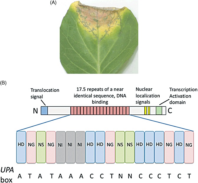
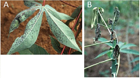
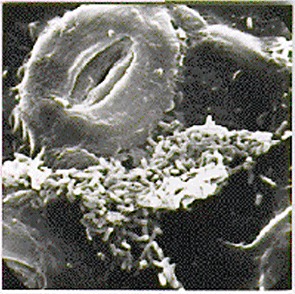
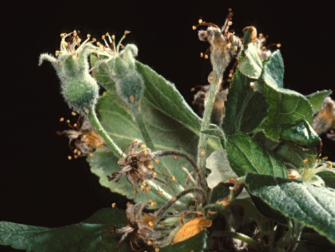
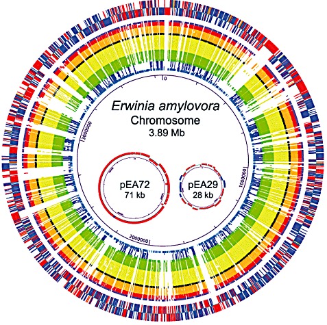
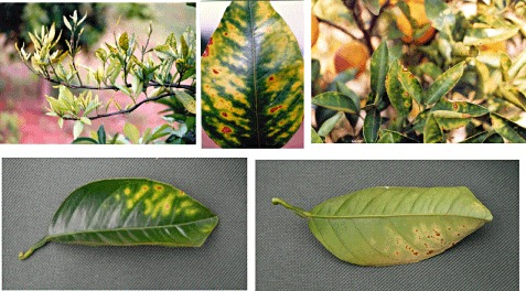
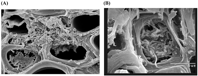
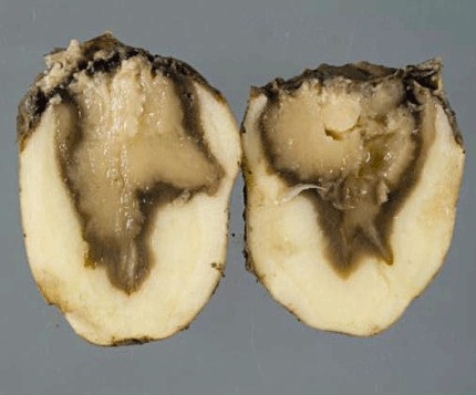
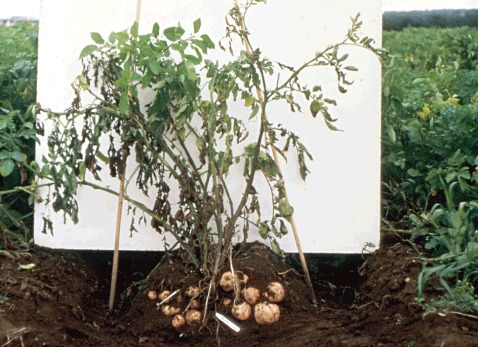
Many plant bacteriologists, if not all, feel that their particular microbe should appear in any list of the most important bacterial plant pathogens. However, to our knowledge, no such list exists. The aim of this review was to survey all bacterial pathologists with an association with the journal Molecular Plant Pathology and ask them to nominate the bacterial pathogens they would place in a 'Top 10' based on scientific/economic importance. The survey generated 458 votes from the international community, and allowed the construction of a Top 10 bacterial plant pathogen list. The list includes, in rank order: (1) Pseudomonas syringae pathovars; (2) Ralstonia solanacearum; (3) Agrobacterium tumefaciens; (4) Xanthomonas oryzae pv. oryzae; (5) Xanthomonas campestris pathovars; (6) Xanthomonas axonopodis pathovars; (7) Erwinia amylovora; (8) Xylella fastidiosa; (9) Dickeya (dadantii and solani); (10) Pectobacterium carotovorum (and Pectobacterium atrosepticum). Bacteria garnering honourable mentions for just missing out on the Top 10 include Clavibacter michiganensis (michiganensis and sepedonicus), Pseudomonas savastanoi and Candidatus Liberibacter asiaticus. This review article presents a short section on each bacterium in the Top 10 list and its importance, with the intention of initiating discussion and debate amongst the plant bacteriology community, as well as laying down a benchmark. It will be interesting to see, in future years, how perceptions change and which bacterial pathogens enter and leave the Top 10.


Figures

Figure 1
Figure 1
The type III secretion system (T3SS) of Pseudomonas syringae pv. tomato. (A) Putative basal body of the T3SS released from membrane preparations after growth in hrp inducing medium. The arrow marks the attachment point of the Hrp pilus. Bar, 25 nm. (B) False colour image of the Hrp pilus gold labelled with antibodies to the subunit protein HrpA, emerging from the bacterial surface. Bar, 50 nm. Both images kindly provided by Ian Brown (University of Kent).
Figure 2
Figure 2
Ralstonia solanacearum (A, photograph J. Vasse) and disease wilting symptoms on tomato (B) with bacteria oozing from the vascular system after stem section (C).
Figure 3
Figure 3
A crown gall on cherry trunk caused by Agrobacterium tumefaciens.
Figure 4
Figure 4
Wild‐type tomato plant developing crown gall tumours (left) and Agrobacterium tumefaciens‐resistant transgenic tomato plant generated by A. tumefaciens‐mediated genetic transformation (right) illustrate two important aspects of A. tumefaciens: one as a pathogen and another as a tool for genetic engineering (reproduced with permission from Escobar et al., 2001).
Figure 5
Figure 5
Visualization of Xanthomonas oryzae pv. oryzae (Xoo) in rice plants. (A, B) Transverse leaf sections of rice infected with Xoo strain PXO99 expressing the green fluorescence of rice cultivar TP309 (susceptible) (A) and TP309‐XA21 (resistant) (B). Images were observed with excitation from 450 to 490 nm and emitted light collected at 520 nm at 40× magnification using a Zeiss Axiophot fluorescence microscope, 12 days after inoculation. The bars in (A) and (B) represent 50 µm. (C) Scanning electron micrograph of Xoo cells in the xylem vessel of a rice leaf. (D) Close‐up of Xoo‐infected rice leaf. Bacterial cells fill the xylem vessels and ooze out at hydathodes, forming beads or strands of exudate on the leaf surface, a characteristic sign of the disease. Photographs in (A) and (B) courtesy of S. W. Han (reprinted from BMC Microbiol. 2008; 8: 164). Photograph in (C) courtesy of J. Leach (reprinted from Mol. Plant Pathol. 2006; 7(5): 303–324). Photograph in (D) courtesy of the Bureau of Rice Research and Development, Thailand (http://www.brrd.in.th).
Figure 6
Figure 6
(A) Black rot disease symptoms on cabbage caused by Xanthomonas campestris pv. campestris, showing the characteristic blackening of the leaf veins (image kindly provided by Sarah Schatschneider and Karsten Niehaus, University of Bielefeld). (B) Domain architecture of the AvrBs3 effector showing the variations at positions 12 and 13 in the repeats and the nucleotides recognized in the consensus UPA (upregulated by AvrBs3) box (see Boch and Bonas, 2010).
Figure 7
Figure 7
Bacterial blight symptoms caused by Xanthomonas axonopodis pv. manihotis: (A) angular leaf spots (Courtesy of V. Verdier, IRD Montpellier, France); (B) leaf wilting (courtesy of B. Boher, IRD Montpellier, France).
Figure 8
Figure 8
Scanning electron microscopy showing a large amount of bacteria near the stomata (Courtesy of V. Verdier, IRD Montpellier, France).
Figure 9
Figure 9
Xanthomonas axonopodis pv. manihotis in xylem vessels (courtesy of B. Boher, IRD Montpellier, France).
Figure 10
Figure 10
Apple blossom cluster infected by Erwinia amylovora.
Figure 11
Figure 11
Circular representation of the genome of Erwinia amylovora strain ATCC 49946 (Ea273) and comparison with related genomes. The figure and legend were provided courtesy of Bryan S. Biehl and Nicole T. Perna (University of Wisconsin, MI, USA), and Ana Maria Bocsanczy and Steven V. Beer (Cornell University, Ithaca, NY, USA).
Figure 12
Figure 12
Symptoms of citrus variegated chlorosis in leaves and plant of sweet orange (photograph Marcos A. Machado).
Figure 13
Figure 13
(A, B) Biofilm of Xylella fastidiosa blocking the xylem vessels of sweet orange tree. Photographs in (A) by E.W. Kitajima (Escola Superior de Agricultura Luis de Queiróz, USP, Piracicaba, SP, Brazil) and in (B) by J.O. Lima (Citrulima Viveiros, São João da Boa Vista, SP, Brazil) and Marcos A. Machado.
Figure 14
Figure 14
Artemis screenshot showing reciprocal best hit analysis of coding sequences (CDS) between Pectobacterium atrosepticum (top) and Dickeya dadantii 3937 (bottom). Coloured lines represent orthologues; red, same orientation; blue, opposite orientation.
Figure 15
Figure 15
Potato tuber rot caused by ‘Dickeya solani’. Fera crown copyright.
Figure 16
Figure 16
Dickeya solani’ expressing green fluorescent protein (GFP) on potato roots (courtesy of J. van der Wolf, Plant Research International, Wageningen, the Netherlands).
Figure 17
Figure 17
Blackleg disease of potato caused by Pectobacterium atrosepticum. Apparently healthy mother tubers can be seen, but stem rotting is also clear.
Figure 18
Figure 18
Identification of Pectobacterium mutants affected in potato plant virulence (stem inoculation assays). Left, wild‐type; others, reduced virulence.

Similar articles

Cited by

References

    1. Antunez‐Lamas, M. , Cabrera, E. , Lopez‐Solanilla, E. , Solano, R. , Gonzalez‐Melendi, P. , Chico, J.M. , Toth, I.K. , Birch, P.R.J. , Pritchard, L. , Liu, H. and Rodriguez‐Palenzuela, P. (2009) Bacterial chemoattraction towards jasmonate plays a role in the entry of Dickeya dadantii through wounded tissues. Mol. Microbiol. 74, 662–671. - PubMed
    1. Arnold, D. , Lovell, H. , Jackson, R.W. and Mansfield, J.W. (2011) Pseudomonas syringae pv. phaseolicola: from ‘has bean’ to supermodel. Mol. Plant Pathol. 12, 617–627. - PMC - PubMed
    1. Babujee, L. , Venkatesh, B. , Yamazaki, A. and Tsuyumu, S. (2007) Proteomic analysis of the carbonate insoluble outer membrane fraction of the soft‐rot pathogen Dickeya dadantii (syn. Erwinia chrysanthemi) strain 3937. J. Proteome Res. 6, 62–69. - PubMed
    1. Baltrus, D.A. , Nishimura, M.T. , Romanchuk, A. , Chang, J.H. , Mukhtar, M.S. , Cherkis, K. , Roach, J. , Grant, S.R. , Jones, C.D. and Dangl, J.L. (2011) Dynamic evolution of pathogenicity revealed by sequencing and comparative genomics of 19 Pseudomonas syringae isolates. PLoS Pathog. 7, e1002132. Epub: 1 July 2011. - PMC - PubMed
    1. Barnard, A. , Bowden, S. , Burr, T. , Corbett, M. , Coulthurst, S. , Monson, R. and Salmond, G.P.C. (2007) Quorum sensing, virulence and secondary metabolite production in plant soft rotting bacteria. Proc. R. Soc. London, Ser. B: Biol. Sci. 362, 1165–1183. - PMC - PubMed