. 2012 Jun 15;11(12):2337-47.
doi: 10.4161/cc.20810. Epub 2012 Jun 15.

Timeless preserves telomere length by promoting efficient DNA replication through human telomeres

Affiliations

Timeless preserves telomere length by promoting efficient DNA replication through human telomeres

Adam R Leman et al. Cell Cycle. .

Abstract

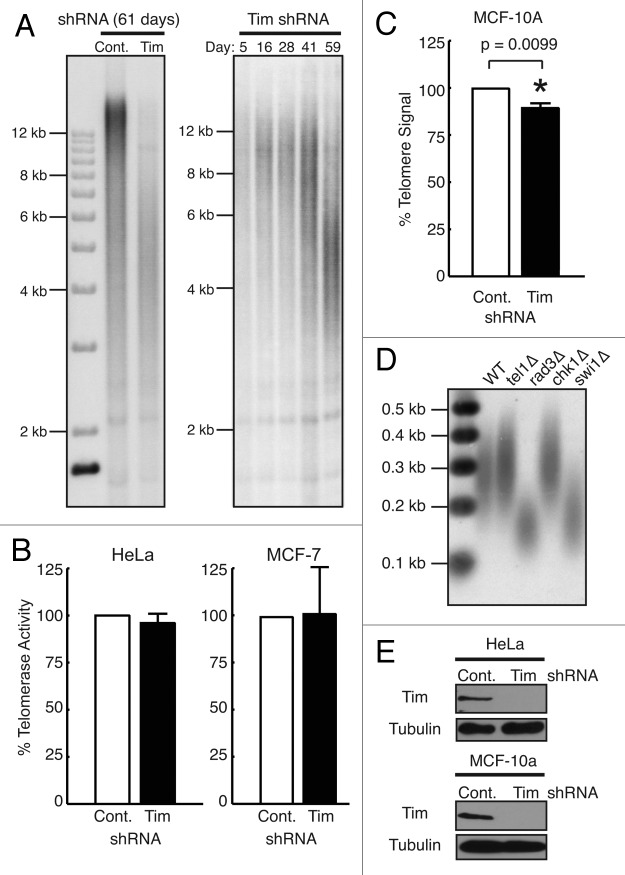
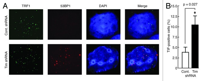
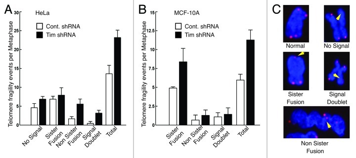
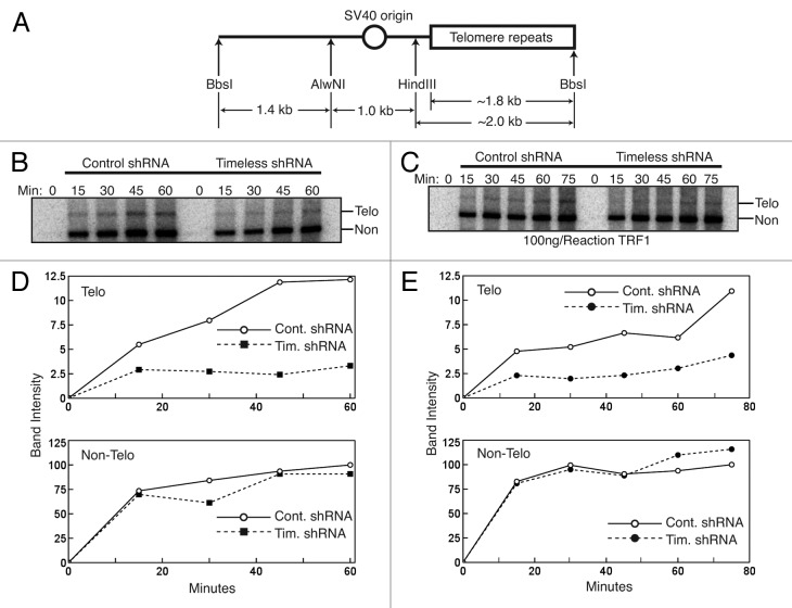
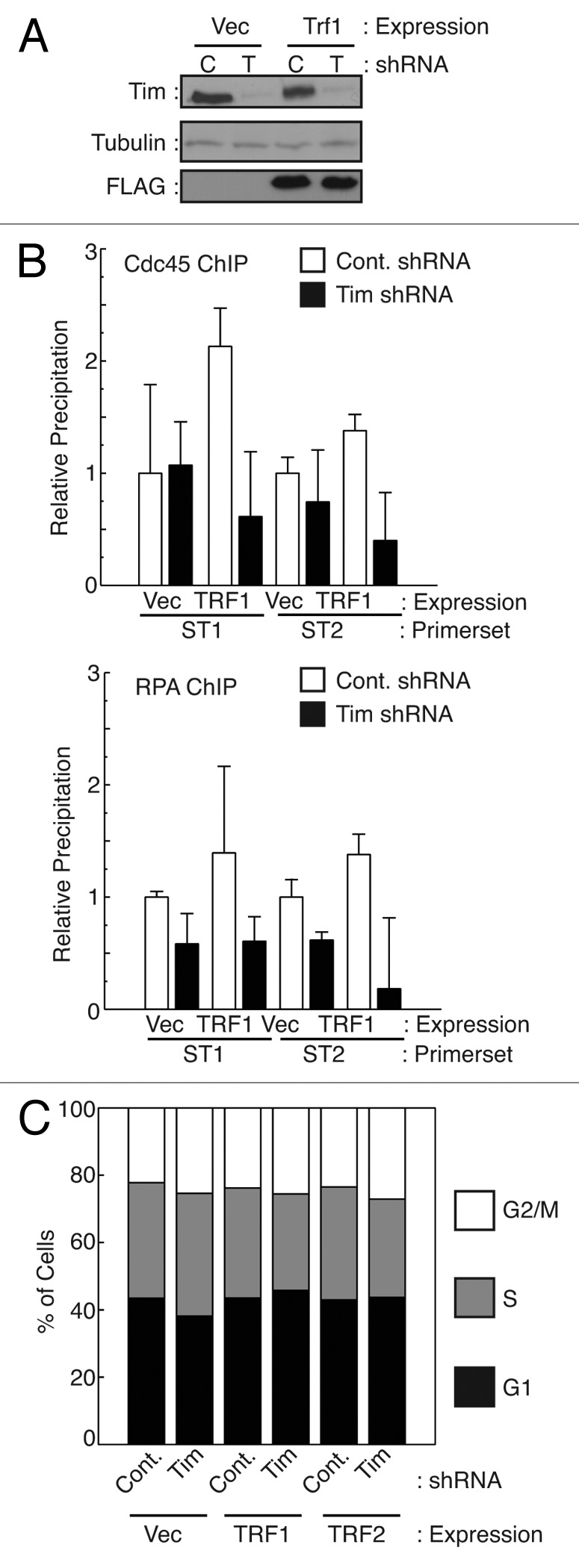
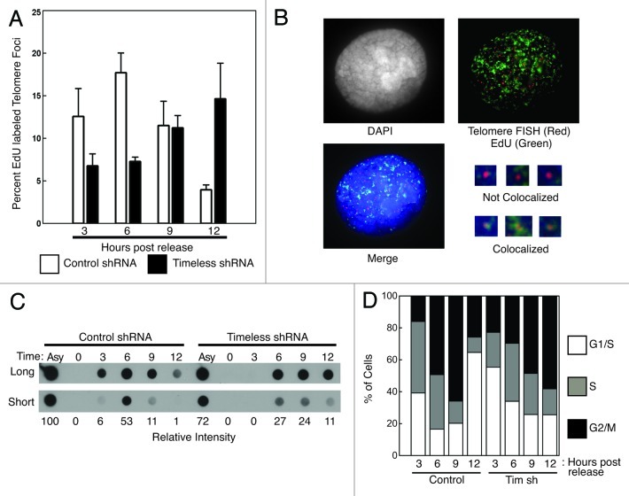
A variety of telomere protection programs are utilized to preserve telomere structure. However, the complex nature of telomere maintenance remains elusive. The Timeless protein associates with the replication fork and is thought to support efficient progression of the replication fork through natural impediments, including replication fork block sites. However, the mechanism by which Timeless regulates such genomic regions is not understood. Here, we report the role of Timeless in telomere length maintenance. We demonstrate that Timeless depletion leads to telomere shortening in human cells. This length maintenance is independent of telomerase, and Timeless depletion causes increased levels of DNA damage, leading to telomere aberrations. We also show that Timeless is associated with Shelterin components TRF1 and TRF2. Timeless depletion slows telomere replication in vitro, and Timeless-depleted cells fail to maintain TRF1-mediated accumulation of replisome components at telomeric regions. Furthermore, telomere replication undergoes a dramatic delay in Timeless-depleted cells. These results suggest that Timeless functions together with TRF1 to prevent fork collapse at telomere repeat DNA and ensure stable maintenance of telomere length and integrity.


Figures

None
Figure 1. Timeless depletion leads to loss of telomeric DNA. (A) Telomere restriction fragment (TRF) Southern blotting analysis in HeLa cells. HeLa cells were infected by lentivirus expressing control or Timeless shRNA and continuously cultured under selection for the indicated days. Genomic DNA was prepared, digested with RsaI and HinfI, and processed for TRF Southern blot analyses using a radiolabeled telomere-specific probe. RsaI and HinfI are frequent cutters that digest genomic DNA, but do not target telomere repeats, generating a broad telomere hybridization signal ranging from 2 kb to 20 kb in HeLa cells. (B) Quantitative telomerase activity assay. Telomerase activity in Timeless shRNA cells is shown as a value relative to the activity in control shRNA cells. Cell line is indicated on each graph. Data are from at least three independent experiments and error bars represent standard deviations. (C) Telomere Flow-FISH assay of MCF-10A cells expressing the indicated shRNA. Six days post infection, cells were subjected to Telomere Flow-FISH using a FITC-conjugated telomere-specific probe, analyzing telomere signal in G1 cells. Error bars correspond to standard deviations obtained from three independent experiments. *The P-value determined by paired Student’s t-test is indicated. (D) Telomere restriction analysis of S. pombe cells. Genomic DNA prepared from the indicated cells was digested by ApaI and processed for Southern blot using a telomere probe. The ApaI site is located 30–40 bp away from telomeric repeat sequences of S. pombe chromosome termini, generating a ~300 bp telomere hybridization signal in the wild-type (WT) strain. (E) western blotting analysis of Timeless (Tim) protein in HeLa and MCF-10A cells expressing the indicated shRNA are shown, confirming knockdown of Timeless protein. Tubulin was used as a loading control.
None
Figure 2. Timeless-depleted cells have telomere specific DNA damage. (A) Telomere dysfunction-induced foci (TIF) assay of MCF-10A cells expressing the indicated shRNA. Cells were in situ-extracted with Triton X-100. Colocalization of 53BP1 and TRF1 was determined using anti-53BP1 (red) and anti-TRF1 (green) antibodies. DNA was co-stained with DAPI (4’, 6’-diamidino-2-phenylindole). The merged images of 53BP1, TRF1 and DNA are shown. (B) Quantification of cells containing TIFs (colocalization of 53BP1 and TRF1). Cells with four or more TIFs were scored as TIF positive (n > 100 nuclei; Error bars correspond to standard deviations obtained from three independent experiments). *Denotes P-value determined by paired Student’s t-test.
None
Figure 3. Timeless-depleted cells have telomere aberrations. (A, B) Frequency of metaphase telomere phenotypes observed in HeLa (A) or MCF-10A (B) cells expressing control or Timeless shRNA. Metaphase spreads were hybridized with a telomere-PNA probe. n > 100 metaphase spreads; Error bars correspond to standard deviations obtained from three independent experiments. (C) Representative images of a normal metaphase chromosome and telomere abnormalities are shown.
None
Figure 4. Timeless interacts with TRF1 and TRF2. (A) Timeless associates with TRF1 and TRF2. 293T cells were transfected with the indicated plasmids to express Timeless or Tipin. 48 h later, immunoprecipitation (FLAG-IP) was performed on cell extracts with the anti-FLAG beads. Associated proteins were probed with anti-TRF1 and anti-TRF2 antibodies. (B) 293T cells were transfected with indicated plasmids to express Shelterin components. 48 h later, immunoprecipitation (FLAG-IP) was performed using anti-FLAG beads. Immunoprecipitates were probed with indicated antibodies to determine interaction between Timeless and Shelterin components. (C) The indicated GST-fused recombinant proteins were incubated with the Timeless protein for 45 min. GST-fusion proteins were precipitated using glutathione Sepharose beads and analyzed by western blotting using the anti-Timeless antibody (Timeless WB). Recombinant Timeless and GST-fusion proteins were stained by Colloidal blue staining. WCE, whole cell extract; FLAG-IP, immunoprecipitated fraction; WB, western blotting. Representative results of repeat experiments are shown.
None
Figure 5. Timeless depletion slows replication of telomere, but not nonrepetitive DNA templates in vitro. (A) Schematic representation of the replication template, pT2AG3 plasmid linearized with BbsI enzyme. The largest fragment (right) contains ~1.8 kb of telomere repeats. (B and C) Autoradiography of radiolabeled replication products from in vitro SV40-mediated DNA replication reactions using the indicated cell extracts. Reactions were stopped at the indicated times and samples were purified, digested, and separated by agarose gel electrophoresis. In (B) no exogenous human protein added. In (C), recombinant TRF1 protein added. (D and E) Quantification of the replication products, at the indicated times, as shown above (in B and C, respectively). Top Panels are telomere DNA products, and bottom Panels are non-telomere DNA products. Representative results of repeat experiments are shown.
None
Figure 6. Timeless downregulation reduces the level of replisome association at telomeres. (A) Western analysis of cells used in ChIP experiments. (B) HeLa cells expressing the indicated shRNA (C: Control; T: Timeless) were transfected with control vector (Vec), pcDNA3-TRF1-FL (TRF1). Cells were processed for ChIP using antibodies against Cdc45 or RPA. Precipitated DNA recovered from antibody-conjugated beads was used to monitor the association of Cdc45 or RPA using primers targeting different sub-telomeric (TTAGGG)n repeats (ST1 and ST2). TRF1 overexpression results in an increase in replisome association at sub-telomeres, which was abrogated by Timeless depletion. Relative DNA precipitation from each sample to control precipitation (set to 1) is shown for each experiment. Data are from at least three independent experiments, and error bars represent standard deviations. (C) Cell cycle analysis of cells used in B was performed using flow cytometry.
None
Figure 7. Telomere replication is profoundly delayed when Timeless is depleted. (A) Timeless depletion caused a profound delay in telomere replication timing. HeLa cells expressing control or Timeless shRNA were synchronized by thymidine at the beginning of S-phase and released. Cells were incubated with EdU for 30 min prior to the time point and processed for EdU detection and telomere-FISH (telomere-PNA probe). Foci containing both EdU and telomere signals were scored as telomere replication foci. Percentages of telomere replication signals over total telomere signals were obtained. Data were obtained from three independent experiments, and error bars represent standard deviations. At least 400 foci were counted for each experiment. (B) Representative image of stained cells with replicating (colocalized EdU and telomere signals) and non-replicating (not colocalized) telomere foci are shown. (C) Timeless depletion caused a mild delay in bulk BrdU incorporation. To assess the rate of DNA replication, cells used in A were also incubated with BrdU for 30 min at the indicated times after the release from a double thymidine block. Genomic DNA was fixed on nitrocellulose membrane to monitor the incorporation of BrdU by western blotting using an anti-BrdU antibody. Relative intensities of signals are shown below. Asy: Asynchronous cells. (D) Cell cycle analysis of cells used for telomere replication assays in (A and B).

Comment in

Similar articles

Cited by

References

    1. Olovnikov AM. [Principle of marginotomy in template synthesis of polynucleotides] Dokl Akad Nauk SSSR. 1971;201:1496–9. - PubMed
    1. Olovnikov AM. A theory of marginotomy. The incomplete copying of template margin in enzymic synthesis of polynucleotides and biological significance of the phenomenon. J Theor Biol. 1973;41:181–90. doi: 10.1016/0022-5193(73)90198-7. - DOI - PubMed
    1. Watson JD. Origin of concatemeric T7 DNA. Nat New Biol. 1972;239:197–201. - PubMed
    1. Bryan TM, Englezou A, Gupta J, Bacchetti S, Reddel RR. Telomere elongation in immortal human cells without detectable telomerase activity. EMBO J. 1995;14:4240–8. - PMC - PubMed
    1. Greider CW, Blackburn EH. The telomere terminal transferase of Tetrahymena is a ribonucleoprotein enzyme with two kinds of primer specificity. Cell. 1987;51:887–98. doi: 10.1016/0092-8674(87)90576-9. - DOI - PubMed

Publication types

MeSH terms

Substances