Review
. 2012 Sep;20(9):411-8.
doi: 10.1016/j.tim.2012.05.002. Epub 2012 Jun 4.

The ParA/MinD family puts things in their place

Affiliations
Review

The ParA/MinD family puts things in their place

Joe Lutkenhaus. Trends Microbiol. 2012 Sep.

Abstract

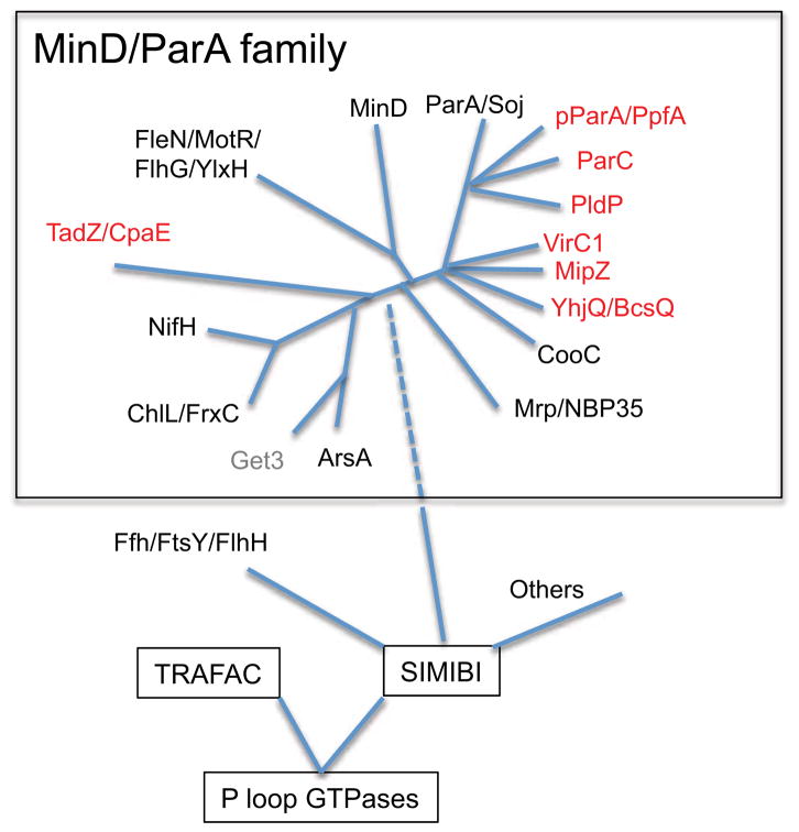
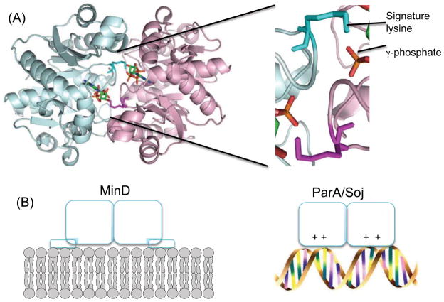
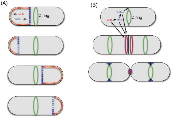
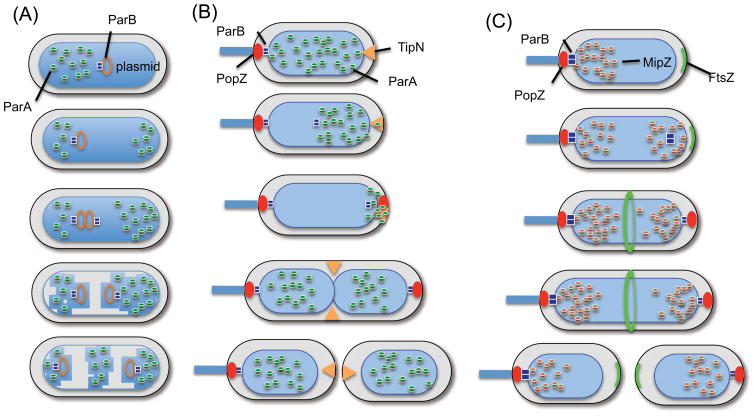
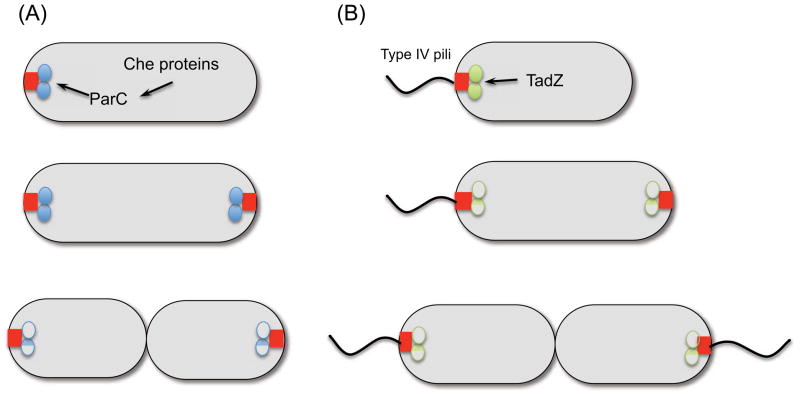
Bacteria must segregate their DNA and position a septum to grow and divide. In many bacteria, MinD is involved in spatial regulation of the cytokinetic Z ring, and ParAs are involved in chromosome and plasmid segregation. The use of the MinD/ParA family to provide positional information for spatial organization continues to expand with the recognition that orphan ParAs are required for segregating cytoplasmic protein clusters and the polar localization of chemotaxis proteins, conjugative transfer machinery, type IV pili, and cellulose synthesis. Also, some bacteria lacking MinD use orphan ParAs to regulate cell division. Positioning of MinD/ParA proteins is either due to self-organization on a surface or reliance on a landmark protein that functions as a molecular beacon.


Figures

Figure 1
Figure 1
Phylogenetic tree displaying the position of MinD/ParA family members. The MinD/ParA family is a part of the SIMIBI class of P loop GTPases. Leipe et al. (6) recognized 8 subfamilies of the MinD/ParA/Mrp family (names in black). Get3 is closely related to ArsA. More recently recognized members are colored red. This diagram was based upon Leipe et al. (6) with additional information obtained from Perez-Cheeks et al. (11) and Ringgaard et al. (36).
Figure 2
Figure 2
MinD/ParA proteins undergo ATP dependent dimerization to bind to surfaces and other proteins. (a) MinD dimer (PDB 3Q9L), with ATP (phosphates in orange) and signature lysines highlighted. The expanded view on the right shows the signature lysine in proximity to the γ-phosphate of ATP bound to other subunit. (b) ATP dependent dimerization of MinD and ParA lead to binding to surfaces. The MinD dimer binds to membranes through a C-terminal amphipathic helix and the ParA/Soj dimer binds nonspecifically to DNA through positive charged residues. Although binding to different surfaces, the orientation of the proteins on the surface is the same.
Figure 3
Figure 3
Localization behavior of MinD. MinD contributes to Z ring positioning in E. coli (a) and B. subtilis (b). In E. coli MinD and MinE oscillate between the poles to prevent Z ring assembly away from midcell. In B. subtilis DivIVA localizes to the incipient septum and recruits MinJ which recruits MinD and MinC. The decorated DivIVA ring is stable as the septum constricts. After the cells split, the Min proteins are released and DivIVA reorganizes.
Figure 4
Figure 4
Pattern formation involving ParA/MipZ and nonspecific DNA binding. ParA promotes the segregation of plasmids and cytoplasmic protein clusters (a) and the origin of chromosomes (b). (a) ParB bound to the parS site located on a plasmid displaces ParA that is bound nonspecifically to DNA. In some cases the cargo is not a plasmid but a cytoplasmic protein cluster (carboxysomes or chemotaxis clusters). (b) During chromosome segregation in C. crescentus the origin region is tethered to the polarity protein PopZ at the pole. Following duplication, one of the ParBs follows the receding ParA. The released ParA is maintained at the pole by TipN until late in the cell cyle when it is released and it spreads over the nucleoid. (c) MipZ, a distinct ParA protein, forms a bipolar gradient on the nucleoid to regulate the position of the Z ring in C. crescentus. The gradient emanates from ParB bound at the origin.
Figure 5
Figure 5
ParC and TadZ are involved in the polar localization of chemotaxis proteins and type IV pili respectively. (a) ParC is related to ParA but does not bind DNA. The polar determinant it binds to is unknown. (b) TadZ is a link between an unknown polar determinant and the type IV pilus machinery.

References

    1. Bi EF, Lutkenhaus J. FtsZ ring structure associated with division in Escherichia coli. Nature. 1991;354:161–4. - PubMed
    1. Webb CD, et al. Bipolar localization of the replication origin regions of chromosomes in vegetative and sporulating cells of B. subtilis. Cell. 1997;88:667–74. - PubMed
    1. Gordon GS, et al. Chromosome and low copy plasmid segregation in E. coli: visual evidence for distinct mechanisms. Cell. 1997;90:1113–21. - PubMed
    1. Thompson SR, et al. The positioning of cytoplasmic protein clusters in bacteria. Proc Natl Acad Sci U S A. 2006;103:8209–14. - PMC - PubMed
    1. Savage DF, et al. Spatially ordered dynamics of the bacterial carbon fixation machinery. Science. 2010;327:1258–61. - PubMed

MeSH terms

Substances