

Structural validation of oral mucosal tissue using optical coherence tomography
- PMID: 22673083
- PMCID: PMC3414779
- DOI: 10.1186/1758-3284-4-29
Structural validation of oral mucosal tissue using optical coherence tomography
Expression of concern in
-
Comment: Head and Neck Oncology.BMC Med. 2014 Feb 5;12:24. doi: 10.1186/1741-7015-12-24. BMC Med. 2014. PMID: 24499430 Free PMC article. Review.
Abstract
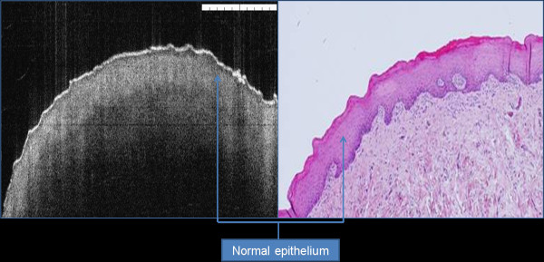
Background: Optical coherence tomography (OCT) is a non-invasive optical technology using near-infrared light to produce cross-sectional tissue images with lateral resolution.
Objectives: The overall aims of this study was to generate a bank of normative and pathological OCT data of the oral tissues to allow identification of cellular structures of normal and pathological processes with the aim to create a diagnostic algorithm which can be used in the early detection of oral disorders.
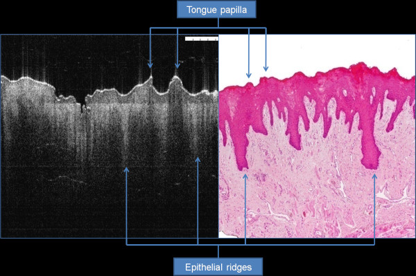
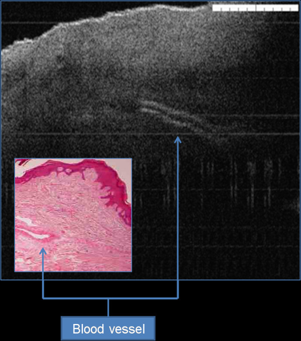
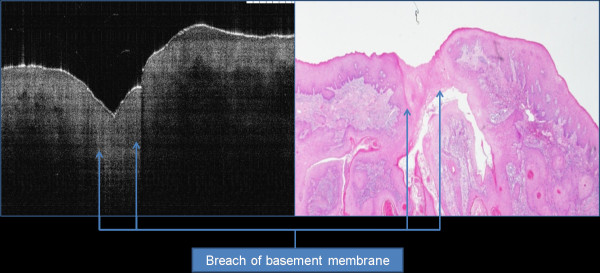
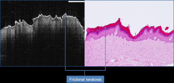
Material and methods: Seventy-three patients with 78 suspicious oral lesions were referred for further management to the UCLH Head and Neck Centre, London. The entire cohort had their lesions surgically biopsied (incisional or excisional). The immediate ex vivo phase involved scanning the specimens using optical coherence tomography. The specimens were then processed by a histopathologist. Five tissue structures were evaluated as part of this study, including: keratin cell layer, epithelial layer, basement membrane, lamina propria and other microanatomical structures. Two independent assessors (clinician and pathologist trained to use OCT) assessed the OCT images and were asked to comment on the cellular structures and changes involving the five tissue structures in non-blind fashion.
Results: Correct identification of the keratin cell layer and its structural changes was achieved in 87% of the cohort; for the epithelial layer it reached 93.5%, and 94% for the basement membrane. Microanatomical structures identification was 64% for blood vessels, 58% for salivary gland ducts and 89% for rete pegs. The agreement was "good" between the clinician and the pathologist. OCT was able to differential normal from pathological tissue and pathological tissue of different entities in this immediate ex vivo study. Unfortunately, OCT provided inadequate cellular and subcellular information to enable the grading of oral premalignant disorders.
Conclusion: This study enabled the creation of OCT bank of normal and pathological oral tissues. The pathological changes identified using OCT enabled differentiation between normal and pathological tissues, and identification of different tissue pathologies. Further studies are required to assess the accuracy of OCT in identification of various pathological processes involving the oral tissues.
Figures
References
-
- Upile T, Jerjes W, Sterenborg HJ, El-Naggar AK, Sandison A, Witjes MJ, Biel MA, Bigio I, Wong BJ, Gillenwater A, MacRobert AJ, Robinson DJ, Betz CS, Stepp H, Bolotine L, McKenzie G, Mosse CA, Barr H, Chen Z, Berg K, D’Cruz AK, Stone N, Kendall C, Fisher S, Leunig A, Olivo M, Richards-Kortum R, Soo KC, Bagnato V, Choo-Smith LP, Svanberg K, Tan IB, Wilson BC, Wolfsen H, Yodh AG, Hopper C. Head & neck optical diagnostics: vision of the future of surgery. Head Neck Oncol. 2009 Jul 13;1:25. doi: 10.1186/1758-3284-1-25. - DOI - PMC - PubMed
-
- Upile T, Jerjes WK, Sterenborg HJ, Wong BJ, El-Naggar AK, Ilgner JF, Sandison A, Witjes MJ, Biel MA, van Veen R, Hamdoon Z, Gillenwater A, Mosse CA, Robinson DJ, Betz CS, Stepp H, Bolotine L, McKenzie G, Barr H, Chen Z, Berg K, D’Cruz AK, Sudhoff H, Stone N, Kendall C, Fisher S, MacRobert AJ, Leunig A, Olivo M, Richards-Kortum R, Soo KC, Bagnato V, Choo-Smith LP, Svanberg K, Tan IB, Wilson BC, Wolfsen H, Bigio I, Yodh AG, Hopper C. At the frontiers of surgery: review. Head Neck Oncol. 2011 Feb 9;3(1):7. doi: 10.1186/1758-3284-3-7. - DOI - PMC - PubMed
-
- Jerjes WK, Upile T, Wong BJ, Betz CS, Sterenborg HJ, Witjes MJ, Berg K, van Veen R, Biel MA, El-Naggar AK, Mosse CA, Olivo M, Richards-Kortum R, Robinson DJ, Rosen J, Yodh AG, Kendall C, Ilgner JF, Amelink A, Bagnato V, Barr H, Bolotine L, Bigio I, Chen Z, Choo-Smith LP, D’Cruz AK, Gillenwater A, Leunig A, MacRobert AJ, McKenzie G, Sandison A, Soo KC, Stepp H, Stone N, Svanberg K, Tan IB, Wilson BC, Wolfsen H, Hopper C. The future of medical diagnostics: review paper. Head Neck Oncol. 2011 Aug 23;3:38. doi: 10.1186/1758-3284-3-38. - DOI - PMC - PubMed
-
- Wong BJ, Jackson RP, Guo S, Ridgway JM, Mahmood U, Su J, Shibuya TY, Crumley RL, Gu M, Armstrong WB, Chen Z. In vivo optical coherence tomography of the human larynx: normative and benign pathology in 82 patients. Laryngoscope. 2005 Nov;115(11):1904–1911. doi: 10.1097/01.MLG.0000181465.17744.BE. - DOI - PubMed
MeSH terms
LinkOut - more resources
Full Text Sources
