

A dimerized urea-based inhibitor of the prostate-specific membrane antigen for 68Ga-PET imaging of prostate cancer
- PMID: 22673157
- PMCID: PMC3502552
- DOI: 10.1186/2191-219X-2-23
A dimerized urea-based inhibitor of the prostate-specific membrane antigen for 68Ga-PET imaging of prostate cancer
Abstract
Background: Alternative positron-emission tomography (PET) probes like labeled inhibitors of the prostate-specific membrane antigen (PSMA) are of emerging clinical impact as they show the ability to image small lesions of recurrent prostate cancer. Here, the dimerization of the pharmacophore Glu-ureido-Lys via the 68Ga chelator N,N'-bis[2-hydroxy-5-(carboxyethyl)benzyl]ethylenediamine-N,N'-diacetic acid (HBED-CC) was investigated to further improve the binding characteristics and pharmacokinetics.
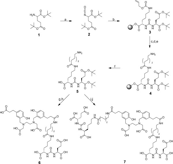
Methods: The peptidomimetic structures were synthesized by solid-phase chemistry, and the resulting products were coupled with the respective 2,3,5,6-tetrafluorophenol esters of HBED-CC to form the monomeric reference and the dimeric Glu-ureido-Lys derivative. The binding properties were analyzed in competitive binding, internalization, and cell surface retention experiments. PET images and biodistribution data were obtained 1 h after injection in BALB/c nu/nu mice bearing LNCaP tumor xenografts.
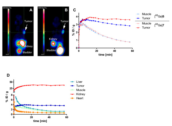
Results: Cell binding data revealed significant better binding properties of the dimer (IC50 = 3.9 ± 1.8 nM; IC50 (monomer) = 12.1 ± 2.1 nM). The inhibition potency investigated by the enzyme-based NAALADase assay confirmed these results. Specific internalization in LNCaP cells was demonstrated for both, the monomer and dimer. As shown by efflux measurements, the dimeric compound was more effectively retained on the cell surface, resulting in advanced in vivo properties (T/BMonomer = 9.2; T/BDimer = 26.5).
Conclusions: The dimeric [68Ga]7 is a promising imaging agent for PSMA-expressing tumors as it shows higher tumor uptake while observing more favorable background clearance. As compared to the respective monomer, the higher affinity and prolonged tumor retention additionally represent promising features and warrant further evaluation regarding 68Ga-PET imaging of PSMA expression.
Figures


References
-
- Andriole GL, Crawford ED, Grubb RL 3rd, Buys SS, Chia D, Church TR, Fouad MN, Gelmann EP, Kvale PA, Reding DJ, Weissfeld JL, Yokochi LA, O'Brien B, Clapp JD, Rathmell JM, Riley TL, Hayes RB, Kramer BS, Izmirlian G, Miller AB, Pinsky PF, Prorok PC, Gohagan JK, Berg CD. Mortality results from a randomized prostate-cancer screening trial. N Engl J Med. 2009;360:1310–1319. doi: 10.1056/NEJMoa0810696. - DOI - PMC - PubMed
-
- Vees H, Buchegger F, Albrecht S, Khan H, Husarik D, Zaidi H, Soloviev D, Hany TF, Miralbell R. 18 F-choline and/or 11 C-acetate positron emission tomography: detection of residual or progressive subclinical disease at very low prostate-specific antigen values (<1 ng/mL) after radical prostatectomy. BJU Int. 2007;99:1415–1420. doi: 10.1111/j.1464-410X.2007.06772.x. - DOI - PubMed
-
- DeGrado TR, Coleman RE, Wang S, Baldwin SW, Orr MD, Robertson CN, Polascik TJ, Price DT. Synthesis and evaluation of 18 F-labeled choline as an oncologic tracer for positron emission tomography: initial findings in prostate cancer. Cancer Res. 2001;61:110–117. - PubMed
-
- Hara T, Kosaka N, Kishi H. PET imaging of prostate cancer using carbon-11-choline. J Nucl Med. 1998;39:990–995. - PubMed
LinkOut - more resources
Full Text Sources
Other Literature Sources
Research Materials
Miscellaneous
