. 2012 Aug;153(8):3701-12.
doi: 10.1210/en.2012-1020. Epub 2012 Jun 6.

Proinflammatory actions of glucocorticoids: glucocorticoids and TNFα coregulate gene expression in vitro and in vivo

Affiliations

Proinflammatory actions of glucocorticoids: glucocorticoids and TNFα coregulate gene expression in vitro and in vivo

Erica A Lannan et al. Endocrinology. 2012 Aug.

Abstract

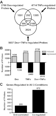
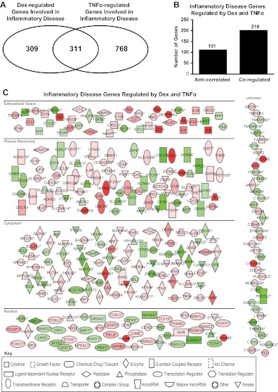
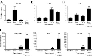
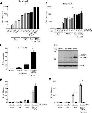
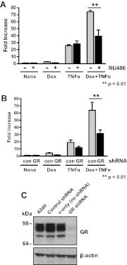
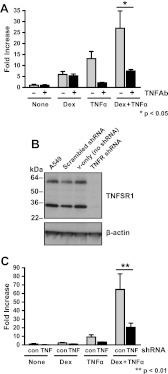
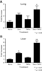
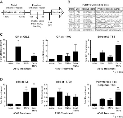
Synthetic glucocorticoids are widely used for treatment of many inflammatory diseases. However, long-term glucocorticoid treatment can cause a variety of negative side effects. A genome-wide microarray analysis was performed in human lung A549 cells to identify genes regulated by both the antiinflammatory steroid dexamethasone (Dex) and the proinflammatory cytokine TNFα. Unexpectedly, we discovered that numerous genes were coregulated by treatment with both Dex and TNFα. We evaluated the mechanism of coregulation of one of these genes, serpinA3 (α-1 antichymotrypsin), a secreted, acute phase protein strongly associated with numerous inflammatory diseases. Up-regulation of serpinA3 requires the presence of both the glucocorticoid receptor and TNFα soluble receptor 1. Treatment with Dex or TNFα resulted in a 10- to 25-fold increase of serpinA3 mRNA, whereas coadministration of Dex and TNFα led to a synergistic increase in serpinA3 mRNA. The naturally occurring glucocorticoid, cortisol, also resulted in a synergistic increase in serpinA3 mRNA levels in A549 cells. Furthermore, in vivo treatment of C57BL/6 mice with Dex and TNFα resulted in coregulation of serpinA3 mRNA levels in both lung and liver tissues. Finally, chromatin immunoprecipitation analyses suggest that glucocorticoid receptor binding to the serpinA3 transcriptional start site can be enhanced by the combination of Dex plus TNFα treatment of A549 cells. These studies demonstrate that glucocorticoids and proinflammatory compounds can coregulate genes associated with human disease. This discovery may underlie the basis of some of the adverse effects associated with long-term glucocorticoid therapy.


Figures

Fig. 1.
Fig. 1.
Identification and classification of Dex- and TNFα-regulated genes. A, Venn diagram of regulated transcripts indicating the overlap between transcripts regulated by the different treatments. B, Bar histogram of up- and down-regulated transcripts for each treatment. C, The 877 transcripts coregulated by Dex, TNFα, and Dex + TNFα were grouped based on their pattern of expression and classified as coregulated or anticorrelated.
Fig. 2.
Fig. 2.
IPA of regulated genes. Genes regulated under the indicated conditions were subjected to IPA to predict potential effects on cellular functions. Genes regulated in inflammatory disorders were selected for further analysis. A, Venn diagram of Dex- or TNFα-regulated genes involved in inflammatory disorders indicating the overlap between the genes regulated by either of these compounds. B, Inflammatory disease genes regulated by Dex and TNFα were classified as coregulated or anticorrelated. C, Diagram listing of inflammatory genes regulated by Dex and TNFα. Green represents up-regulated genes, and red represents down-regulated genes; the brighter the color, the stronger the regulation. Each symbol represents a different class of gene products (see legend on figure). Asterisks indicate that multiple identifiers in the dataset file map to a single gene in the Global Molecular Network.
Fig. 3.
Fig. 3.
qRT-PCR validations of regulated genes. Gene expression patterns from the microarray were validated by one-step quantitative RT-PCR. Predeveloped and validated primer/probe sets were purchased for all genes analyzed (Applied Biosystems). A549 cells were treated with 10 nm of Dex, 10 μg/ml TNFα, or treated with both compounds, and RNA was isolated from these cells after 6 h of treatment. Target gene expression levels were normalized to an endogenous control (cyclophilin B) in all experiments, and data were plotted as fold change relative to control (hormone free) mRNA for statistical comparisons. A, Example of a Dex-up-regulated gene (Dusp1). B, Example of a TNFα-up-regulated gene (TLR3). C, Example of a gene regulated in cells treated with both Dex and TNFα (C3). D, Examples of genes coup-regulated by treatment with Dex and TNFα (SAA1, SAA2, and SerpinA3).
Fig. 4.
Fig. 4.
Characterization of serpinA3 regulation. A549 cells were treated with Dex, TNFα, or both Dex and TNFα, and mRNA levels were analyzed by qRT-PCR. All samples were compared with control (none) for statistical analysis. A, Dose response. Cells were treated for 6 h with the indicated concentrations of Dex, TNFα, or both Dex and TNFα. Results are plotted in log scale. B, Time course of treatment. Cells were treated with Dex, TNFα, or both Dex and TNFα for the indicated lengths of time. Results are plotted in log scale. C, Cells were treated for 6 h with the indicated concentrations of cortisol instead of Dex. D, Western blot analysis of AACT (serpinA3) protein levels. β-Actin is used as a loading control. E, Cells were pretreated with either vehicle or cycloheximide, followed by treatment with Dex, TNFα, or both Dex and TNFα. serpinA3 mRNA levels were analyzed by qRT-PCR. F, Cells were pretreated with either vehicle or actinomycin D, followed by treatment with Dex, TNFα, or both Dex and TNFα. serpinA3 mRNA levels were analyzed by qRT-PCR.
Fig. 5.
Fig. 5.
GR-dependent mechanism of action. A, A549 cells were treated with Dex, TNFα, or both Dex + TNFα for 6 h in conjunction with GR antagonist RU486, and mRNA was isolated. serpinA3 mRNA levels were analyzed by qRT-PCR. RU486 and control samples were compared in each treatment for the statistical analysis. B, A549 cells were treated with lentivirus containing shRNA for GR knockdown or control scrambled shRNA. A549 cells were treated with Dex, TNFα, or both Dex + TNFα for 6 h, and mRNA was isolated. serpinA3 mRNA levels were analyzed by qRT-PCR. Control and GR shRNA were compared in each treatment for the statistical analysis. C, Western blot analysis was performed on cells treated with lentivirus containing scrambled shRNA, vector only, or GR-specific shRNA to confirm reduction in GR protein levels. Con, Control.
Fig. 6.
Fig. 6.
TNFR-dependent mechanism of action. A, A549 cells were treated with Dex, TNFα, or both Dex and TNFα in conjunction with TNFα antibody, and mRNA was isolated. serpinA3 mRNA levels were analyzed by qRT-PCR. TNFα antibody and control samples were compared in each treatment for the statistical analysis. B, Western blot analysis was performed on cells treated with lentivirus containing scrambled shRNA, vector only, or TNFR-specific shRNA to confirm reduction in TNFSR1A protein levels. C, A549 cells were treated with lentivirus containing shRNA for TNFR knockdown or control scrambled shRNA. A549 cells were treated with Dex, TNFα, or both Dex + TNFα for 6 h, and mRNA was isolated. serpinA3 mRNA levels were analyzed by qRT-PCR. Control and TNFR shRNA were compared in each treatment for the statistical analysis.
Fig. 7.
Fig. 7.
In vivo responses to Dex and TNFα treatment. Mice were injected ip with 1 mg/kg Dex, 60 μg/kg TNFα, or both Dex and TNFα, and after 6 h, animals were killed, and mRNA was isolated from the indicated tissues. serpinA3 mRNA levels were analyzed by qRT-PCR. A, Lung. B, Liver.
Fig. 8.
Fig. 8.
ChIP of serpinA3 promoter. A549 cells were treated with 10 nm of Dex, 10 μg/ml TNFα, or both Dex and TNFα for 90 min. ChIP assays were performed on the indicated regions. A, Schematic of GR promoter region used for ChIP assay. B, Schematic of putative GR binding sites. C, ChIP assay using GR at the promoter region of Gilz, a site 1700 bp upstream of the SerpinA3 transcriptional start site, and at the transcriptional start site of serpinA3. D, ChIP assay using the p65 subunit of nuclear factor κB (NF-κB) at the promoter region of IL-8, at a site 1700 bp upsteam of the SerpinA3 transcriptional start site, and polymerase II at the transcriptional start site of SerpinA3. TSS, Transcriptional start site.

Similar articles

Cited by

References

    1. Revollo JR, Cidlowski JA. 2009. Mechanisms generating diversity in glucocorticoid receptor signaling. Ann NY Acad Sci 1179:167–178 - PubMed
    1. Baschant U, Tuckermann J. 2010. The role of the glucocorticoid receptor in inflammation and immunity. J Steroid Biochem Mol Biol 120:69–75 - PubMed
    1. Magnussen H, Watz H. 2009. Systemic inflammation in chronic obstructive pulmonary disease and asthma: relation with comorbidities. Proc Am Thorac Soc 6:648–651 - PubMed
    1. Ross KR, Chmiel JF, Konstan MW. 2009. The role of inhaled corticosteroids in the management of cystic fibrosis. Paediatr Drugs 11:101–113 - PMC - PubMed
    1. Lane NE, Yao W. 2010. Glucocorticoid-induced bone fragility. Ann NY Acad Sci 1192:81–83 - PMC - PubMed

Publication types

MeSH terms