. 2012 Sep;108(5):1453-72.
doi: 10.1152/jn.00274.2012. Epub 2012 Jun 6.

Force encoding in stick insect legs delineates a reference frame for motor control

Affiliations

Force encoding in stick insect legs delineates a reference frame for motor control

Sasha N Zill et al. J Neurophysiol. 2012 Sep.

Abstract

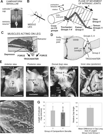
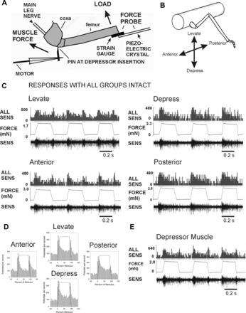
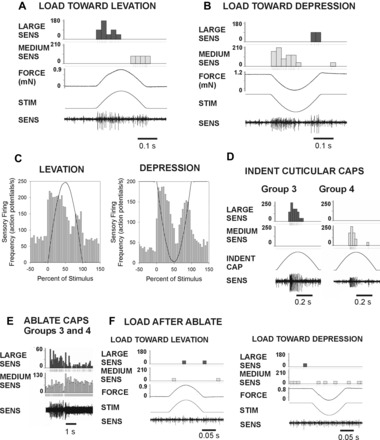
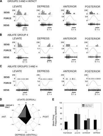
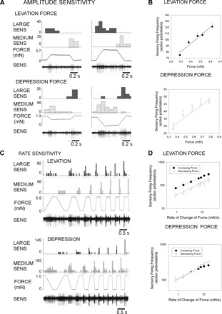
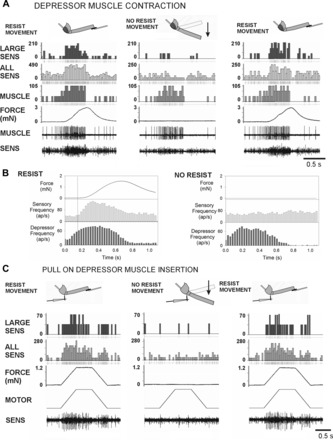
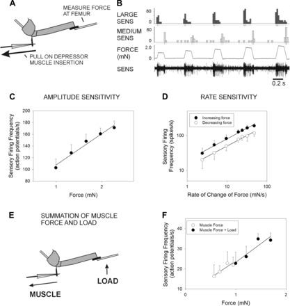
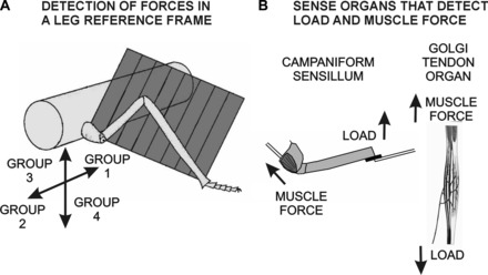
The regulation of forces is integral to motor control. However, it is unclear how information from sense organs that detect forces at individual muscles or joints is incorporated into a frame of reference for motor control. Campaniform sensilla are receptors that monitor forces by cuticular strains. We studied how loads and muscle forces are encoded by trochanteral campaniform sensilla in stick insects. Forces were applied to the middle leg to emulate loading and/or muscle contractions. Selective sensory ablations limited activities recorded in the main leg nerve to specific receptor groups. The trochanteral campaniform sensilla consist of four discrete groups. We found that the dorsal groups (Groups 3 and 4) encoded force increases and decreases in the plane of movement of the coxo-trochanteral joint. Group 3 receptors discharged to increases in dorsal loading and decreases in ventral load. Group 4 showed the reverse directional sensitivities. Vigorous, directional responses also occurred to contractions of the trochanteral depressor muscle and to forces applied at the muscle insertion. All sensory discharges encoded the amplitude and rate of loading or muscle force. Stimulation of the receptors produced reflex effects in the depressor motoneurons that could reverse in sign during active movements. These data, in conjunction with findings of previous studies, support a model in which the trochanteral receptors function as an array that can detect forces in all directions relative to the intrinsic plane of leg movement. The array could provide requisite information about forces and simplify the control and adaptation of posture and walking.


Figures

Fig. 1.
Fig. 1.
Structure of leg and trochanteral campaniform sensilla. A: campaniform sensilla monitor strains in the exoskeleton through the dendritic insertion of the sensory neuron to a cuticular cap. B: drawing of middle leg and locations of campaniform sensilla (redrawn after Cruse and Bartling 1995). The leg is segmented and attached to the body at the basal segment (coxa). The body-coxa joint has considerable freedom of movement. The more distal leg articulations are intrinsic hinge joints that act in a single plane. The distribution of groups of campaniform sensilla is not uniform. No groups are found on the coxa, many groups (Groups 1–4) are concentrated on the trochanter, and single groups are found on the distal segments or subsegments (Groups 5–11). CS, campaniform sensilla. C: interior view of the thorax and muscles that act on the leg (redrawn after Marquardt 1940). The largest muscles that move the leg (retractor, protractor, levator, and depressor muscles) are located in the thorax and insert onto the coxa (Cx) and trochanter (Tr). D: drawing of anterior side of the coxa and trochanter. The groups of campaniform sensilla are located distal to the coxo-trochanteral joint condyle and are therefore in the leg plane. E: reconstructions of confocal images of the exoskeleton. i: Anterior view of trochanter—Group 2 sensilla are located on the anterior side of the trochanter opposite the anterior condyle of the coxo-trochanteral hinge joint. ii: Posterior view—Group 1 receptors are situated on the posterior side and have a similar orientation relative to the posterior joint condyle. iii: Dorsal view—Groups 3 and 4 are located on the dorsal side of the trochanter adjacent to the joint with the femur. iv: Interior view of the posterior half of the trochanter—a thick internal projection (buttress) reinforces the trochanter adjacent to the insertion of the trochanteral depressor muscle. F: scanning electron micrograph showing cuticular caps of Groups 3 and 4. G: histograms of sizes and orientation of sensilla. Each group contains cuticular caps of different sizes (left), and the largest caps of Groups 3 and 4 are mutually perpendicular (right) [mean difference between Groups 3 and 4 = 93.8 ± 13.4° (SD), N = 8].
Fig. 2.
Fig. 2.
Preparation and responses of sensilla with all groups intact. A: preparation. Loads were applied to the femur with a force probe linked to a piezoelectric crystal. The probe contained strain gauges and was used to monitor forces in all experiments. Movement of the leg was resisted by a pin inserted into a small hole adjacent to the depressor muscle insertion. In experiments emulating the effects of muscle contractions, forces were applied to the pin via a computer-controlled linear motor. Sensory activities were recorded in the main leg nerve (nervus cruris) proximal to the coxa; the nerve was crushed close to the mesothoracic ganglion. B: orientation of forces relative to the leg plane. Loads were applied in different directions (levation, depression, anterior, posterior) by rotating the micromanipulator that held the force probe. C: recordings during load application with all groups of campaniform sensilla intact (other receptors ablated). Applying loads via the probe with ramp and hold functions produced sensory discharges in all directions. D: mean sensory discharges: histograms plotting the mean sensory discharges during application of forces in each direction. Discharges occurred during the rising phase that rapidly adapted to lower levels during the hold phase. Prominent phasic discharges occurred to decreasing forces in all directions. E: response to forces applied at the depressor muscle insertion. Intense firing at similar amplitudes occurred when movements of the femur were blocked by the force probe. Bursts were also present during force decreases.
Fig. 3.
Fig. 3.
Responses of trochanteral campaniform sensilla Groups 3 and 4. All sensilla except Groups 3 and 4 were first ablated. Forces of small amplitude were applied via the probe and resisted by the pin. A: force in the direction of levation produced large-amplitude spikes during force increase and a burst of smaller amplitude during force decrement. B: loads applied toward levation produced the opposite pattern, with smaller units firing during the rise phase and larger units during the force decline. C: histogram of mean firing during levation and depression. Firing rose rapidly after the stimulus onset. Discharges during force decreases were consistently observed in both levation and depression. D: indentation of cuticular caps. Mechanical stimulation of the caps of individual Group 3 and Group 4 sensilla produced unitary sensory discharges of amplitude similar to those seen following force application to the leg. E: ablation of Groups 3 and 4 produced intense discharges of similar amplitudes. F: sensory bursts did not occur upon application of forces toward levation or depression after cap ablation.
Fig. 4.
Fig. 4.
Selective ablation of sensilla and directional tuning. The specificity of the responses of the dorsal groups was tested by applying forces in different directions and by selective ablation of Group 4. A: Groups 3 and 4 Intact: recordings of tests in which half-sine waveforms were applied to the probe in the joint plane (levation, depression) and perpendicular to the plane of joint movement (anterior, posterior). Discharges were obtained to both force increases and decreases in all directions. B: Ablate Group 4 sensilla: specific components of the responses were eliminated by ablation of Group 4. Discharges still occurred to force increases in the direction of levation and anterior force application and to force decreases in depression and to posterior forces. Firing to other directions was eliminated. C: Ablate Groups 3 and 4: subsequent ablation of Group 3 caps eliminated all bursting upon force application or release. D: polar plot of mean discharge rates during force increases before and after ablation of Group 4. With both groups intact (dark area), the highest rates of firing occurred in the plane of joint movement. After ablation of Group 4 (lighter gray), only discharges to levation and anterior force persisted. F: histogram of mean firing frequencies and standard deviations. Ablation eliminates responses to depression and posterior forces.
Fig. 5.
Fig. 5.
Rate and amplitude sensitivity. A: amplitude sensitivity: recordings of responses to ramp and hold waveforms of variable amplitude (constant rate of change). Firing of sensilla of the largest extracellular amplitude was sustained in tests applied toward levation, while firing of receptors of smaller amplitude was increased to forces applied toward depression. B: plots of mean discharge frequencies during the hold phase show that sensilla could encode force amplitude. C: sensory activities during application of ramp and hold waveforms of variable rate of change but constant amplitude. All sensilla showed strong rate sensitivities to both force increases and decreases. D: plots of mean discharge frequencies during forces increases and decreases. Receptors effectively encoded the rate of change of force. When forces were applied toward levation, sensory response frequencies to force increases were higher than force decreases. Responses were more closely equivalent for forces applied toward depression.
Fig. 6.
Fig. 6.
Responses to spontaneous depressor muscle contractions and forces applied at the muscle insertion. A: spontaneous contractions of the trochanteral depressor muscle in a preparation in which all sensilla were ablated except for Groups 3 and 4. Activities of the slow depressor motoneuron were recorded myographically (MUSCLE). Sensory activities (SENS, bottom) were recorded from nervus cruris, which was crushed distal to the depressor motor nerve branch. Muscle contractions were first resisted (RESIST MOVEMENT) by the probe that registered the resultant forces on the femur (FORCE). The probe was then moved to allow the femur to move freely (NO RESIST MOVEMENT) and then returned to the contact with the femur. Firing of sensory units with large extracellular amplitudes occurred when movement was resisted. This firing did not occur when the resistance was removed but returned after the probe again blocked leg movements. B: histograms of sensory discharges during resisted and unresisted muscle contractions. The increase in sensory firing was concurrent with the force increase, not at the time of initiation of muscle bursting. The rise in sensory frequency did not occur when the probe was moved away and the leg moved freely toward depression. ap, Action potential. C: forces imposed at the depressor muscle insertion. Preparation similar to A but the depressor nerve branch was cut, permitting more complete exposure of the nervus cruris and better discrimination of action potentials. Vigorous responses occurred to forces applied at the insertion of the trochanteral depressor muscle. Discrete responses also occurred during force decreases when the ramp rate was sufficiently high. Sensillum did not fire when the probe was removed and the leg moved freely but returned if the probe was repositioned to resist the force.
Fig. 7.
Fig. 7.
Rate and amplitude sensitivity to forces applied at the depressor muscle insertion. A: preparation. Sensory activities were recorded in preparations in which all sensilla were ablated except for Groups 3 and 4. Forces that mimicked depressor contractions were applied through a pin placed at the depressor muscle insertion. Movement was resisted and forces monitored by a probe placed against the femur. B: amplitude sensitivity. Response discharges increased in frequency to increases in the amplitude of the stimulus (same animal as in Fig. 3). C: plot of mean discharge in the hold phase. Sensilla effectively encode the force amplitude. D: plot of mean discharge frequencies to forces applied at different rates. Sensory firing to force increases and decreases show rate sensitivity. E: summation of muscle forces and load. Sensory activity was first recorded when force was applied to the depressor insertion, and the probe was only used to monitor the forces (as in A). The same force was then applied at the depressor, and a small force was also applied simultaneously by activating the piezoelectric crystal that held the force probe. F: plot of sensory discharge to muscle forces and to muscle forces and small loads in a single preparation. The combination of muscle forces and loads acted like a simple summation, and the sensory discharges were merely shifted to a higher range.
Fig. 8.
Fig. 8.
Effects of trochanteral campaniform sensilla on activity in the trochanteral depressor muscle. A: recording of effects of repetitive indentation of the cuticular caps of Group 4 campaniform sensilla on tonic activities of the slow trochanteral depressor motoneuron (Ds). Each mechanical stimulus produced a transient increase in the depressor firing frequency. B: histogram of tests from 3 preparations shows consistent depressor excitation at short latency from Group 4 stimulation. C: effects of stimulation of the cap of a Group 3 sensillum on depressor firing. Depressor firing was completely inhibited by sensillum activation. D: histogram of tests (from the same 3 preparations) shows consistent inhibition following indentation of Group 3 sensilla. E and F: effects of forces applied in the direction of levation in a preparation with only Group 3 and 4 sensilla intact. In preparations showing tonic postural activity, forces applied toward levation produced inhibition of depressor firing (E, top; F, top). Large active muscle contractions could be evoked after stimulation of the abdomen or cercus. Forces applied toward levation then produced excitation of the depressor (E, middle; F, middle). Both the inhibitory and excitatory effects were eliminated by ablation of the trochanteral campaniform sensilla (E, bottom; F, bottom).
Fig. 9.
Fig. 9.
Model of force detection and control. A: frame of reference. Our findings suggest that the trochanteral sensilla are organized to supply information about the vectorial direction of loads relative to the plane of leg movement. Trochanteral Group 3 receptors show the largest discharge to forces applied dorsally in the leg plane, while Group 4 sensilla respond to ventral forces. Previous tests have demonstrated that the Group 2 receptors encode forces in an anterior direction, while Group 1 discharge to posterior forces. Thus the plane of joint movement forms a frame of reference for information about forces acting on the leg. B: campaniform sensilla share properties of other force receptors. Both campaniform sensilla and Golgi tendon organs encode muscle forces. Detection of load depends upon the presence of muscle tensions.

Similar articles

Cited by

References

    1. Akay T. The Role of Sensory Signals for Interjoint Coordination in Stick Insect Legs (Carausius morosus and Cuniculina impigra) (PhD thesis). Cologne, Germany: University of Cologne, 2002
    1. Akay T, Bässler U, Gerharz P, Büschges A. The role of sensory signals from the insect coxa-trochanteral joint in controlling motor activity of the femur-tibia joint. J Neurophysiol 85: 594–604, 2001 - PubMed
    1. Akay T, Büschges A. Load signals assist the generation of movement-dependent reflex reversal in the femur-tibia joint of stick insects. J Neurophysiol 96: 3532–3537, 2006 - PubMed
    1. Akay T, Haehn S, Schmitz J, Büschges A. Signals from load sensors underlie interjoint coordination during stepping movements of the stick insect leg. J Neurophysiol 92: 42–51, 2004 - PubMed
    1. Akay T, Ludwar BC, Göritz ML, Schmitz J, Büschges A. Segment specificity of load signal processing depends on walking direction in the stick insect leg muscle control system. J Neurosci 27: 3285–3294, 2007 - PMC - PubMed

Publication types

LinkOut - more resources