. 2012 Jul 2:9:117.
doi: 10.1186/1742-2094-9-117.

Inhibition of Src kinase activity attenuates amyloid associated microgliosis in a murine model of Alzheimer's disease

Affiliations

Inhibition of Src kinase activity attenuates amyloid associated microgliosis in a murine model of Alzheimer's disease

Gunjan Dhawan et al. J Neuroinflammation. .

Abstract

Background: Microglial activation is an important histologic characteristic of the pathology of Alzheimer's disease (AD). One hypothesis is that amyloid beta (Aβ) peptide serves as a specific stimulus for tyrosine kinase-based microglial activation leading to pro-inflammatory changes that contribute to disease. Therefore, inhibiting Aβ stimulation of microglia may prove to be an important therapeutic strategy for AD.

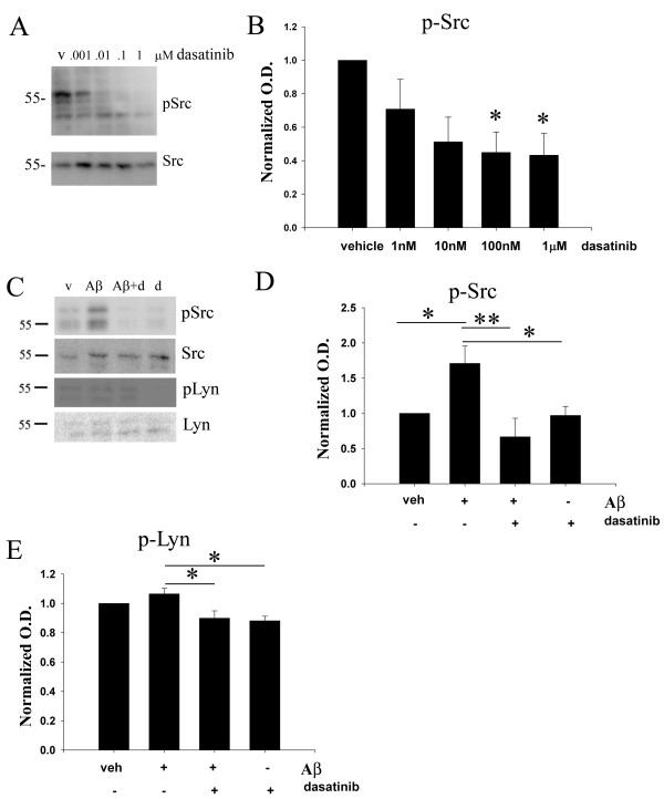
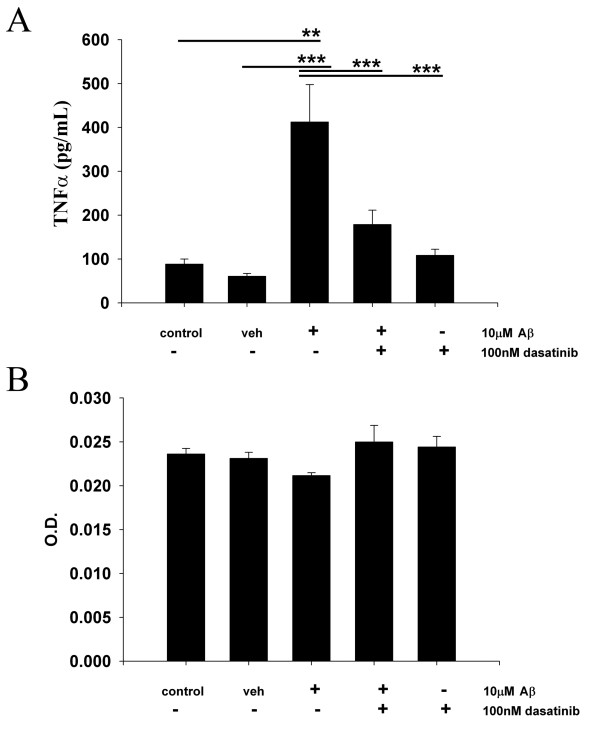
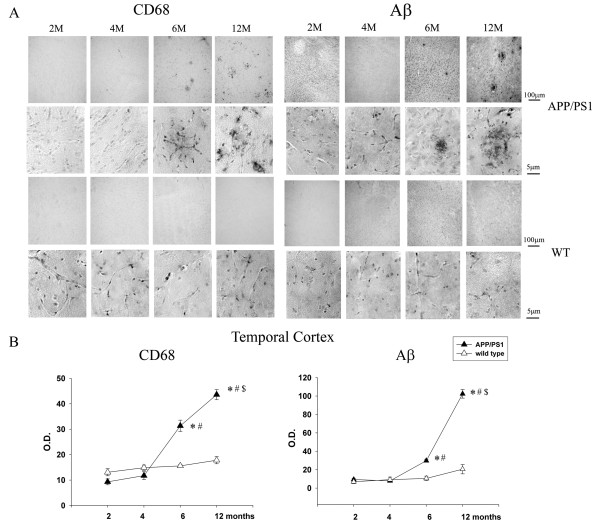
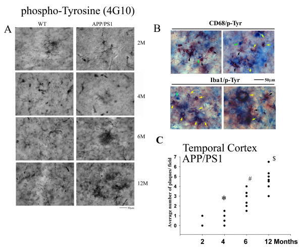
Methods: Primary murine microglia cultures and the murine microglia cell line, BV2, were used for stimulation with fibrillar Aβ1-42. The non-receptor tyrosine kinase inhibitor, dasatinib, was used to treat the cells to determine whether Src family kinase activity was required for the Aβ stimulated signaling response and subsequent increase in TNFα secretion using Western blot analysis and enzyme-linked immunosorbent assay (ELISA), respectively. A histologic longitudinal analysis was performed using an AD transgenic mouse model, APP/PS1, to determine an age at which microglial protein tyrosine kinase levels increased in order to administer dasatinib via mini osmotic pump diffusion. Effects of dasatinib administration on microglial and astroglial activation, protein phosphotyrosine levels, active Src kinase levels, Aβ plaque deposition, and spatial working memory were assessed via immunohistochemistry, Western blot, and T maze analysis.

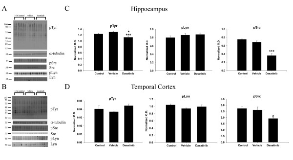
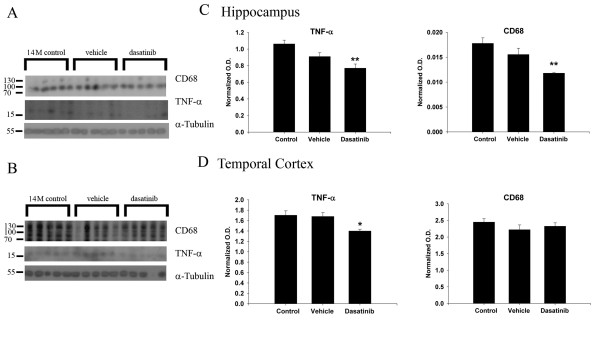
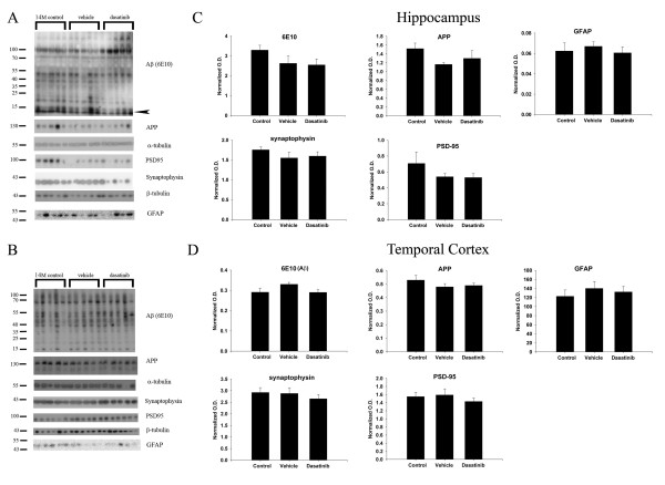
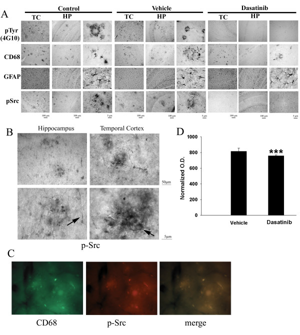
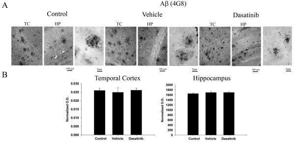
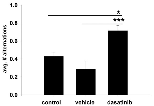
Results: Aβ fibrils stimulated primary murine microglia via a tyrosine kinase pathway involving Src kinase that was attenuated by dasatinib. Dasatinib administration to APP/PS1 mice decreased protein phosphotyrosine, active Src, reactive microglia, and TNFα levels in the hippocampus and temporal cortex. The drug had no effect on GFAP levels, Aβ plaque load, or the related tyrosine kinase, Lyn. These anti-inflammatory changes correlated with improved performance on the T maze test in dasatinib infused animals compared to control animals.

Conclusions: These data suggest that amyloid dependent microgliosis may be Src kinase dependent in vitro and in vivo. This study defines a role for Src kinase in the microgliosis characteristic of diseased brains and suggests that particular tyrosine kinase inhibition may be a valid anti-inflammatory approach to disease. Dasatinib is an FDA-approved drug for treating chronic myeloid leukemia cancer with a reported ability to cross the blood-brain barrier. Therefore, this suggests a novel use for this drug as well as similar acting molecules.


Figures

Figure 1
Figure 1
Dasatinib dose-dependently attenuated active, phospho-Src kinase levels in the BV2 cell line and primary microglia. (A) Microglial BV2 cells were vehicle treated (v), or treated with 1 nM, 10 nM, 100 nM and 1 μM dasatinib for 30 minutes. Cells lysates were resolved by SDS-PAGE and Western blotted using anti-phospho-Src, and Src antibodies. (B) Densitometric analyses of the Western blots were performed normalizing active-Src levels against their respective Src controls and averaging +/−SEM. Blots are representative of six independent experiments. Primary microglia were DMSO vehicle treated (veh), or stimulated for 5 minutes with 10 μM Aβf in the presence or absence of 30-minute pretreatment of 100 nM dasatinib. (C) Cells lysates were Western blotted using anti-pSrc or Src (loading control) antibodies and anti-pLyn or Lyn (loading control) antibodies. A representative blot from six independent experiments is shown. Densitometric analyses of the Western blots was performed normalizing (D) active, phospho-Src levels against Src controls and (E) active, phospho-Lyn levels against Lyn controls and averaging +/−SEM. Percent fold changes in phospho-Src and phospho-Lyn levels were plotted. (*P <0.05 vs. vehicle, dasatinib, **P <0.01 vs. Aβ + dasatinib for pSrc and *P <0.01 vs. Aβ for pLyn).
Figure 2
Figure 2
Dasatinib attenuated Aβ1-42 stimulated-microglial secretion of the proinflammatory cytokine, TNF-α. Primary microglia were unstimulated (control), DMSO vehicle treated (veh), or stimulated for 24 hours with 10 μM Aβf in the presence/absence of 100 nM dasatinib. (A) Media was collected from treated cells and used to quantify changes in TNF-α secretion via ELISA. Secreted values were averaged +/−SEM. (B) After media removal, the treated cells were used to assess cell viability via the MTT reduction assay. Absorbance values (560/650 nm) were averaged+/−SEM. (**P <0.01 from control, ***P <0.001 vs. vehicle, dasatinib and Aβ + dasatinib).
Figure 3
Figure 3
Microgliosis in the APP/PS1 model of AD positively correlated with increased Aβ plaque density with age. Serial coronal sections from 2- , 4- , 6- and 12-month old controls (C57BL/6) and age-matched transgenic animals (APP/PS1) (n = 5 to 6) were immunostained for (A) activated microglia (anti-CD68) and Aβ (anti-4G8 antibody). Representative hemi-sections are shown with select high magnification fields. (B) Immunoreactivity densities from the temporal cortex region for CD68 and 4G8 were quantified, averaged and graphed (+/−SEM) for *P <0.001 from 2- and 4-month APP/PS1 animals, #P <0.001 from 2-, 4-, 6- and12-month wild type animals for CD68, #P <0.001 from 2-, 4- and 6-month wild type mice for 4 G8, and $P <0.001 from 6-month APP/PS1 animals.
Figure 4
Figure 4
Plaque-associated microglial phosphotyrosine immunoreactivity increased with age in APP/PS1 mice. (A) Serial coronal sections from 2- , 4- , 6- and 12-month-old controls (C57BL/6) and age-matched transgenic animals (APP/PS1) (n = 5 to 6) were immunostained with anti-phosphotyrosine antibody (4G10). Representative select high magnification fields from the temporal cortex of hemisections are presented. (B) To validate microglial co-localization of phospho-tyrosine immunoreactivity, 12-month APP/PS1 sections were double-labeled using anti-phosphotyrosine antibodies and VIP as the chromogen (black arrows) and anti-CD68 or Iba1 and Vector Blue as the chromogen (green arrows). Double-labeled cells are indicated with yellow arrows. (C) To quantify immunoreactivity and omit the inclusion of non-microglial phosphotyrosine immunoreactivity, 4G10 positive plaques from APP/PS1 mice were counted from the temporal cortex and averaged and graphed (+/−SEM) for *P <0.001 from two-month APP/PS1 mice, #P <0.001 from two-month and four-month APP/PS1 animals, $P <0.001 from four- and six-month APP/PS1 animals.
Figure 5
Figure 5
Dasatinib infusion reduced protein phospho-tyrosine and active phospho-Src kinase levels in APP/PS1 brains. Vehicle or dasatinib (500 ng/kg/day) was infused subcutaneously into 13-month-old APP/PS1 for 28 days (n = 7). Hippocampus and temporal cortex regions were dissected from the left hemispheres and resolved by 10% SDS-PAGE and Western blotted. (A) Hippocampal and (B) temporal cortex brain lysates from control (14-month APP/PS1), vehicle infused, and dasatinib infused animals were blotted using anti-phosphotyrosine, pTyr (4G10), pSrc, pLyn, α-tubulin, Src, and Lyn antibodies. Optical densities were averaged and graphed (+/−SEM) from blots of (C) hippocampus (*P <0.05 from controls, ***P <0.001 from vehicle) and (D) temporal cortex. (*P <0.05 from controls).
Figure 6
Figure 6
Dasatinib infusion reduced CD68 and TNF-α protein levels in APP/PS1 brains. (A) Hippocampal and (B) temporal cortex brain lysates from control, vehicle, and dasatinib infused animals were Western blotted for activated microglia using anti-CD68 antibody and anti-pro-inflammatory cytokine TNF-α antibody along with anti-α-tubulin as a loading control. Optical densities were averaged and graphed (+/−SEM) from blots of (C) hippocampus (**P <0.01 from controls) and (D) temporal cortex (*P <0.05 from controls).
Figure 7
Figure 7
Dasatinib infusion did not alter levels of APP, Aβ, synaptophysin, PSD95, or GFAP levels in APP/PS1 brains. (A) Hippocampal and (B) temporal cortex brain lysates from control, vehicle, and dasatinib infused animals were used for Western blot analyses using anti-Aβ clone 6E10 antibody, anti-APP, anti-synaptophysin, anti-PSD95, anti-GFAP, anti-α-tubulin and βIII tubulin (loading control) antibodies. Optical densities for each antibody were normalized to tubulin loading controls, averaged, and graphed (+/−SEM) for (C) hippocampus and (D) temporal cortex.
Figure 8
Figure 8
Dasatinib infusion attenuated microgliosis, protein phosphotyrosine and active phospho-Src immunoreactivity with no effect on GFAP immunoreactivity in APP/PS1 brains. Right hemispheres from brains of dasatinib infused, vehicle infused and APP/PS1 controls animals were fixed, serially sectioned and immunostained using (A) anti-phosphotyrosine (4 G10), anti-CD68, anti-GFAP, and anti-phospho-Src (pSrc) antibodies. Representative images from the CA1 region of the hippocampus (HP) as well as the temporal cortex (TC) are shown with select high magnification TC fields. (B) High magnification images of pSrc immunoreactivities from control APP/PS1 temporal cortex and hippocampus demonstrate robust plaque-associated immunoreactivity (arrow) along with vascular immunoreactivity (arrow). (C) Fluorescent double-labeling of control APP/PS1 brains demonstrated clear pSrc (red) immunoreactivity co-localizing with CD68 (green) staining. (D). Relative levels of pSrc immunoreactivity in the temporal cortex were quantified from serial stained sections from vehicle and dasatinib infused brains, averaged (+/−SEM), and graphed (***P <0.001 via student’s t-test).
Figure 9
Figure 9
Dasatinib infusion did not affect Aβ plaque load in the brains of APP/PS1 mice. Serial sections of right hemispheres from brains of dasatinib infused, vehicle infused, and control APP/PS1 mice were immunostained using anti-Aβ antibody (clone 4 G8) and plaque load was quantified. (A) Representative images of the temporal cortex (TC) and hippocampus (HP) from brain hemispheres are shown with select high magnification TC fields. (B) Mean optical density measurements from the CA1 region of the hippocampus as well as the temporal cortex are shown +/− SEM.
Figure 10
Figure 10
Dasatinib infused APP/PS1 mice demonstrated increased spontaneous alternations during T maze testing. Control untreated 14-month APP/PS1 mice, dasatinib infused mice or vehicle infused controls (n = 7) were used for T maze testing on Day 29 following 28 days of drug or vehicle infusion. The mean number of spontaneous alternations +/− SEM per treatment group were graphed. (*P <0.05 vs. control, ***P <0.001 vs. vehicle).

Similar articles

Cited by

References

    1. Akiyama H, Mori H, Saido T, Kondo H, Ikeda K, McGeer PL. Occurrence of the diffuse amyloid beta-protein (Abeta) deposits with numerous Abeta-containing glial cells in the cerebral cortex of patients with Alzheimer's disease. Glia. 1999;25:324–331. doi: 10.1002/(SICI)1098-1136(19990215)25:4<324::AID-GLIA2>3.0.CO;2-5. - DOI - PubMed
    1. Benzing WC, Wujek JR, Ward EK, Shaffer D, Ashe KH, Younkin SG, Brunden KR. Evidence for glial-mediated inflammation in aged APP(SW) transgenic mice. Neurobiol Aging. 1999;20:581–589. doi: 10.1016/S0197-4580(99)00065-2. - DOI - PubMed
    1. Itagaki S, McGeer PL, Akiyama H, Zhu S, Selkoe D. Relationship of microglia and astrocytes to amyloid deposits of Alzheimer disease. J Neuroimmunol. 1989;24:173–182. doi: 10.1016/0165-5728(89)90115-X. - DOI - PubMed
    1. Mattiace LA, Davies P, Dickson DW. Detection of HLA-DR on microglia in the human brain is a function of both clinical and technical factors. Am J Pathol. 1990;136:1101–1114. - PMC - PubMed
    1. Mackenzie IR, Hao C, Munoz DG. Role of microglia in senile plaque formation. Neurobiol Aging. 1995;16:797–804. doi: 10.1016/0197-4580(95)00092-S. - DOI - PubMed

Publication types

MeSH terms