. 2012 Sep 1;303(5):C490-8.
doi: 10.1152/ajpcell.00138.2012. Epub 2012 Jun 6.

The UDP-sugar-sensing P2Y(14) receptor promotes Rho-mediated signaling and chemotaxis in human neutrophils

Affiliations

The UDP-sugar-sensing P2Y(14) receptor promotes Rho-mediated signaling and chemotaxis in human neutrophils

Juliana I Sesma et al. Am J Physiol Cell Physiol. .

Abstract

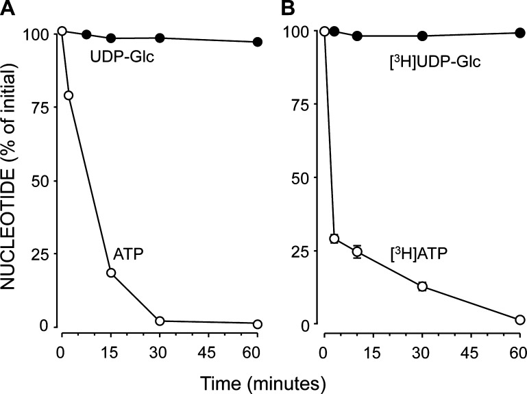
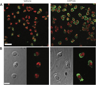
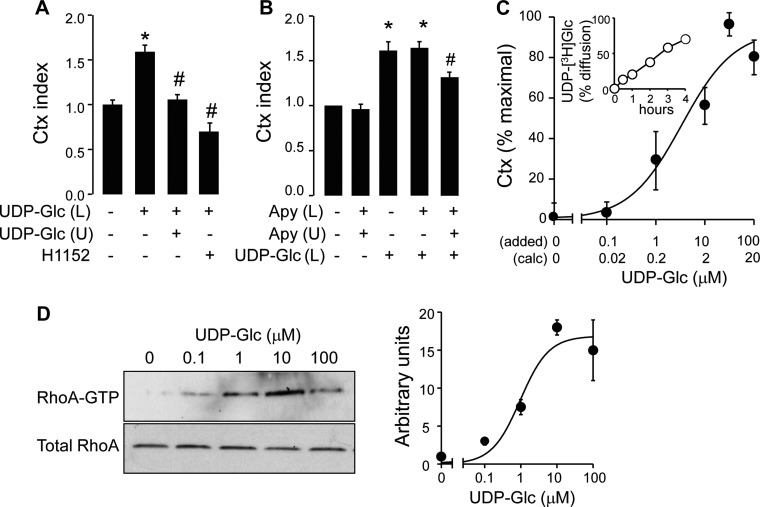
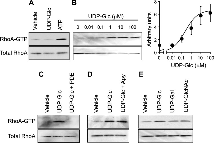
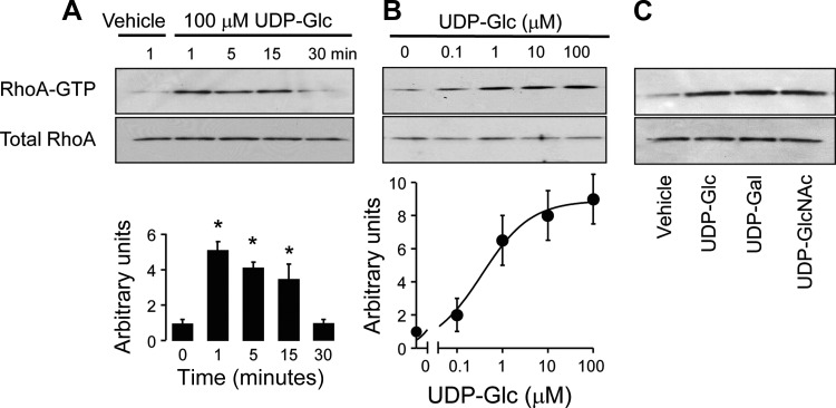
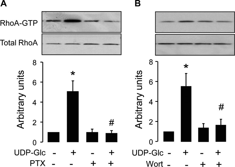
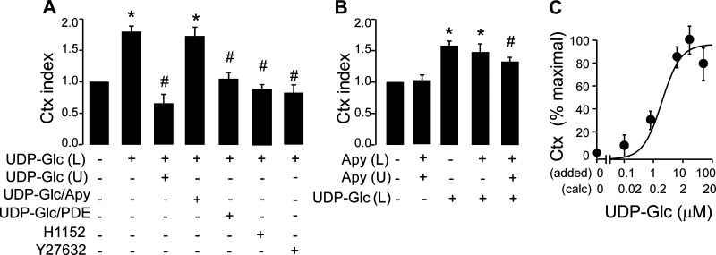
The G(i)-coupled P2Y(14) receptor (P2Y(14)-R) is potently activated by UDP-sugars and UDP. Although P2Y(14)-R mRNA is prominently expressed in circulating neutrophils, the signaling pathways and functional responses associated with this receptor are undefined. In this study, we illustrate that incubation of isolated human neutrophils with UDP-glucose resulted in cytoskeleton rearrangement, change of cell shape, and enhanced cell migration. We also demonstrate that UDP-glucose promotes rapid, robust, and concentration-dependent activation of RhoA in these cells. Ecto-nucleotidases expressed on neutrophils rapidly hydrolyzed extracellular ATP, but incubation with UDP-glucose for up to 1 h resulted in negligible metabolism of the nucleotide-sugar. HL60 human promyelocytic leukemia cells do not express the P2Y(14)-R, but neutrophil differentiation of HL60 cells with DMSO resulted in markedly enhanced P2Y(14)-R expression. Accordingly, UDP-glucose, UDP-galactose, and UDP-N-acetylglucosamine promoted Rho activation in differentiated but not in undifferentiated HL60 cells. Stable expression of recombinant human P2Y(14)-R conferred UDP-sugar-promoted responses to undifferentiated HL60 cells. UDP-glucose-promoted RhoA activation also was accompanied by enhanced cell migration in differentiated HL60 cells, and these responses were blocked by Rho kinase inhibitors. These results support the notion that UDP-glucose is a stable and potent proinflammatory mediator that promotes P2Y(14)-R-mediated neutrophil motility via Rho/Rho kinase activation.


Figures

Fig. 1.
Fig. 1.
Stability of UDP-glucose (UDP-Glc) in human neutrophils. Neutrophils were incubated for the indicated times with 100 μM UDP-glucose or ATP (A) or trace amounts (0.1 μCi) of UDP-[3H]glucose or [3H]ATP (B). Nucleotides were separated and quantified by HPLC. The data are representative of two independent experiments performed in duplicate.
Fig. 2.
Fig. 2.
UDP-glucose induces cell shape changes and cytoskeleton remodeling in human neutrophils. Neutrophils were incubated for 15 min with vehicle or 100 μM UDP-glucose. Cells were fixed and stained with fluorescent phalloidin (actin cytoskeleton) and propidium iodide (nucleus) as described in materials and methods. A: overlay of confocal planes representing actin and nuclear staining of neutrophils. Bar, 25 μm. B: overlay of confocal planes representing actin and nuclear staining and the corresponding differential interference contrast confocal plane (gray images) of neutrophils at high-power magnification. Bar, 10 μm. The images are representative of two separate experiments in which 15–20 fields were randomly analyzed for each condition.
Fig. 3.
Fig. 3.
UDP-glucose promotes chemotaxis (Ctx) and RhoA activation in human neutrophils. A: neutrophil migration in response to vehicle or 100 μM UDP-glucose added to either the lower (L) or the upper (U) compartment of a Boyden chamber. As indicated, neutrophils were preincubated for 15 min with 1 μM H1152. Results are expressed as chemotaxis index (see materials and methods) and are means ± SE from three separate experiments, each one performed in quadruplicate. *,#Significantly different from vehicle and UDP-glucose (lower), respectively, P < 0.05 by 2-way ANOVA. B: neutrophil migration in response to vehicle or 100 μM UDP-glucose added to the lower compartment. Apyrase (Apy; 5 U/ml) was included in the lower and/or upper compartment, as indicated. Results are means ± SE from two separate experiments, each one performed in quadruplicate. *,#Significantly different from vehicle and UDP-glucose without apyrase, respectively, P < 0.05 by 2-way ANOVA. C: concentration-effect relationship for UDP-glucose-promoted neutrophil chemotaxis. The data (means ± SE, n = 4) are representative of two independent experiments. The values on the x-axis indicate the concentration of agonist added to the lower compartment (added), and the concentration of agonist calculated (calc) to reach the upper compartment at the time of measurement (see inset). Inset: time course for the diffusion of 100 μM UDP-[3H]glucose through the filter membrane (means ± SD, n = 3; representative of two experiments). D: RhoA activation was measured in neutrophils that were incubated for 1 min in the presence of the indicated concentrations of UDP-glucose. The data are presented as means ± SE of results from three separate experiments.
Fig. 4.
Fig. 4.
UDP-sugars promote RhoA activation in differentiated HL60 (dHL60) cells. A: RhoA activation was measured in undifferentiated HL60 cells incubated for 1 min with vehicle, 100 μM UDP-glucose, or 100 μM ATP. B: concentration-effect relationship for UDP-glucose-evoked RhoA activation in dHL60 cells. Error bars indicate differences to the mean from two independent experiments. C: the effect of 100 μM UDP-glucose on dHL60 cells was abolished by pretreatment (15 min) of the nucleotide-sugar with 1 U/ml nucleotide-pyrophosphatase [a phosphodiesterase (PDE)]. D: apyrase (1 U/ml, 15 min) had no effect on UDP-glucose-evoked RhoA activation in dHL60 cells. E: RhoA activation was measured in dHL60 cells incubated with vehicle or 100 μM UDP-glucose, UDP-galactose (UDP-Gal), or UDP-N-acetyl-glucosamine (UDP-GlcNAc). All incubations (A–E) were for 1 min.
Fig. 5.
Fig. 5.
P2Y14 receptor (P2Y14-R) confers UDP-sugar-promoted RhoA activation to undifferentiated HL60 cells. A: undifferentiated P2Y14-HL60 cells were incubated for the time indicated with 100 μM UDP-glucose. The data are presented as means ± SE of results from three separate experiments. *Significantly different from vehicle, P < 0.05 by 2-way ANOVA. B: cells were incubated for 1 min with the indicated concentrations of UDP-glucose. The data are presented as means ± SE of results from three separate experiments. C: cells were incubated for 1 min with vehicle or 100 μM of UDP-glucose, UDP-galactose, or UDP-N-acetyl-glucosamine; the results are representative of three experiments.
Fig. 6.
Fig. 6.
Pertussis toxin (PTX) and a phosphoinositide 3-kinase inhibitor abolish P2Y14-R-promoted Rho activation in HL60 cells. dHL60 cells were preincubated overnight with vehicle or 100 ng/ml of pertussis toxin (A) or 15 min with vehicle or 100 nM wortmannin (Wort) (B) and stimulated for 1 min with 100 μM UDP-glucose. The data are presented as means ± SE of results from four separate experiments. *,#Significantly different from vehicle and UDP-glucose, respectively, P < 0.05 by 2-way ANOVA.
Fig. 7.
Fig. 7.
UDP-glucose promotes chemotaxis in dHL60 cells. A: chemotaxis of dHL60 cells was measured in response to vehicle or 100 μM UDP-glucose added either to the lower or to the upper compartment of the Boyden chamber, as indicated. UDP-glucose was preincubated with 1 U/ml of apyrase or nucleotide pyrophosphatase (PDE) for 15 min before addition of the agonist to the lower compartment, and HL60 cells were preincubated with vehicle, 1 μM H1152, or 5 μM Y27632 for 15 min. Results are means ± SE from three separate experiments, each one performed in quadruplicate. *,#Significantly different from vehicle and UDP-glucose (lower), respectively, P < 0.05 by 2-way ANOVA. B: dHL60 cells migration in response to vehicle or 100 μM UDP-glucose added to the lower compartment. Apyrase (5 U/ml) was included in the lower and/or upper compartment, as indicated. Results are means ± SE from three separate experiments, each one performed in sextuplicate. *,#Significantly different from vehicle and UDP-glucose without apyrase, respectively, P < 0.05 by 2-way ANOVA. C: concentration-effect relationship for UDP-glucose-promoted chemotaxis in dHL60 cells; the values on the x-axis indicate the concentration of agonist added to the lower compartment (added), and the concentration of agonist calculated to reach the upper compartment (calc), as in Fig. 3C. The results represent means ± SE from two separate experiments, each performed in quadruplicate.

Comment in

Similar articles

Cited by

References

    1. Alblas J, Ulfman L, Hordijk P, Koenderman L. Activation of Rhoa and ROCK are essential for detachment of migrating leukocytes. Mol Biol Cell 12: 2137– 2145, 2001 - PMC - PubMed
    1. Arase T, Uchida H, Kajitani T, Ono M, Tamaki K, Oda H, Nishikawa S, Kagami M, Nagashima T, Masuda H, Asada H, Yoshimura Y, Maruyama T. The UDP-glucose receptor P2RY14 triggers innate mucosal immunity in the female reproductive tract by inducing IL-8. J Immunol 182: 7074– 7084, 2009 - PubMed
    1. Brautigam VM, Dubyak GR, Crain JM, Watters JJ. The inflammatory effects of UDP-glucose in N9 microglia are not mediated by P2Y14 receptor activation. Purinergic Signal 4: 73– 78, 2008 - PMC - PubMed
    1. Buell G, Michel AD, Lewis C, Collo G, Humphrey PP, Surprenant A. P2X1 receptor activation in HL60 cells. Blood 87: 2659– 2664, 1996 - PubMed
    1. Carter RL, Fricks IP, Barrett MO, Burianek LE, Zhou Y, Ko H, Das A, Jacobson KA, Lazarowski ER, Harden TK. Quantification of Gi-mediated inhibition of adenylyl cyclase activity reveals that UDP is a potent agonist of the human P2Y14 receptor. Mol Pharmacol 76: 1341– 1348, 2009 - PMC - PubMed

Publication types

LinkOut - more resources