. 2012 Jun 6:3:884.
doi: 10.1038/ncomms1893.

Melanoma induction by ultraviolet A but not ultraviolet B radiation requires melanin pigment

Affiliations
Free PMC article

Melanoma induction by ultraviolet A but not ultraviolet B radiation requires melanin pigment

Frances P Noonan et al. Nat Commun. .
Free PMC article

Abstract

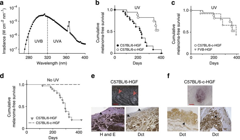
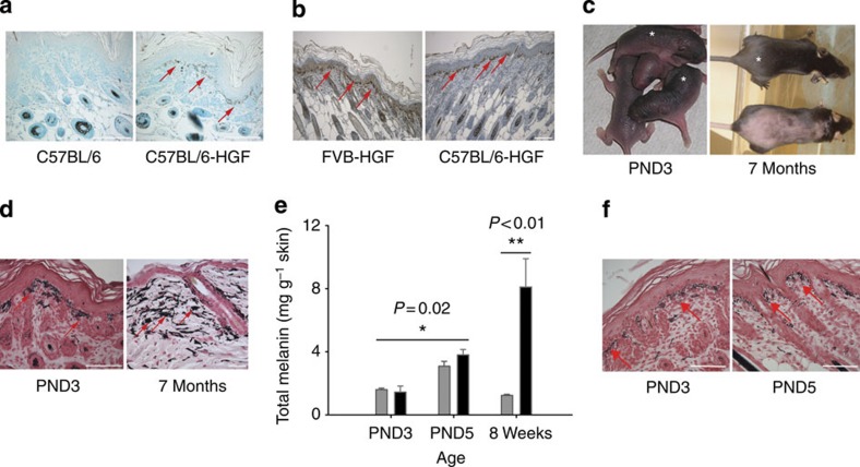
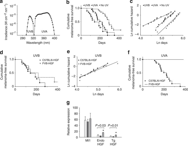
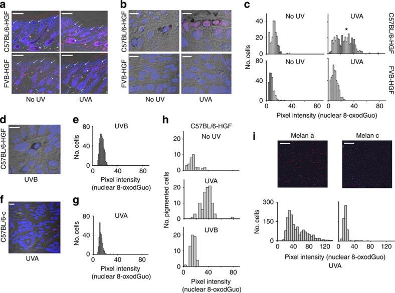
Malignant melanoma of the skin (CMM) is associated with ultraviolet radiation exposure, but the mechanisms and even the wavelengths responsible are unclear. Here we use a mammalian model to investigate melanoma formed in response to precise spectrally defined ultraviolet wavelengths and biologically relevant doses. We show that melanoma induction by ultraviolet A (320-400 nm) requires the presence of melanin pigment and is associated with oxidative DNA damage within melanocytes. In contrast, ultraviolet B radiation (280-320 nm) initiates melanoma in a pigment-independent manner associated with direct ultraviolet B DNA damage. Thus, we identified two ultraviolet wavelength-dependent pathways for the induction of CMM and describe an unexpected and significant role for melanin within the melanocyte in melanomagenesis.


Figures

Figure 1
Figure 1. Melanoma in ultraviolet-irradiated black and albino HGF transgenic mice.
A single melanomagenic dose from F40 sunlamps of 9.5 kJ m−2 (23 SED) of ultraviolet radiation containing approximately 65% ultraviolet B and 34% ultraviolet A was delivered to 3-day-old neonatal black C57BL/6-HGF and albino C57BL/6-c-HGF- tyrosinase-mutant pups, which were followed for melanoma development. (a) Ultraviolet (UV) spectral irradiance of F40 sunlamp broadband source. (b) Melanoma in irradiated black C57BL/6-HGF (black circles) and albino C57BL/6-c-HGF (white circles) mice (P<0.001, log-rank test). (c) Melanoma in irradiated albino C57BL/6-c-HGF mice (white circles) compared with previously reported melanoma in albino FVB-HGF mice irradiated with the same dose of ultraviolet from the same source (white triangles; P=0.9, log-rank test). (d) Melanoma in unirradiated black and albino animals. No spontaneous melanomas occurred in albino C57BL/6-c-HGF mice and spontaneous melanomas arose in black C57BL/6-HGF animals (grey triangles) more slowly than in irradiated mice (P<0.003, log-rank test). (e) Multiple tumours arose in black C57BL/6-HGF mice (red arrows). Pigmented clusters of melanoma cells in the epidermis and at the dermal–epidermal junction (black arrows) were confirmed as melanoma by bleaching and immunohistochemistry for the melanocytic marker Dct. (f) Single tumours that arose in albino C57BL/6-c-HGF mice were invasive into the subcutaneous fat and into the epidermis (black arrows). Red scale bars, 5 mm. White scale bars, 100 μm.
Figure 2
Figure 2. Melanocyte and melanin distribution in unirradiated animals.
(a) Extra-follicular pigmented cells (arrows) are visible in methyl green-stained skin of 3-day-old C57BL/6-HGF transgenic but not C57BL/6 wild-type mice. (b) Comparable numbers of extra-follicular melanocytes (arrows), visualized by immunohistochemistry for Dct, were observed in skin of 3-day-old albino FVB-HGF and black C57BL/6-HGF mice. Scale bars, 100 μm. (c) Pigmentation of C57BL/6-HGF transgenics (asterisk) and C57BL/6 wild-type littermates (unmarked) at 3 days of age (PND3) and of shaved 7 month old C57BL/6-HGF transgenic mouse (asterisk) compared to shaved wild type animal (unmarked). (d) Melanin (black, Fontana stain) in skin of PND3 and 7-month-old C57BL/6-HGF mice is largely confined to melanocytes and is sparse in the epidermis. Melanosomes were visible in neonatal melanocytes (arrows) but adult (7 months) melanocytes were heavily congested with pigment (arrows). Scale bars, 100 μm. (e) Total melanin in C57BL/6 (grey bars) and C57BL/6-HGF transgenic (black bars) mouse skin determined by ESR was similar between genotypes at each neonatal time point (P>0.1, t-test) but increased significantly in both genotypes between PND3 and PND5 (* t-test). Adult HGF transgenic skin (8 weeks) had more than sixfold the melanin of adult wild-type skin (** t-test). Bars represent mean±s.e.m. of 3–5 biological replicates. No melanin was detectable in albino skin. (f) Frequencies of extra-follicular melanocytes (arrows) are similar in 3- and 5-day-old (PND5) neonatal C57BL/6-HGF skin. Fontana stain. Scale bars, 100 μm.
Figure 3
Figure 3. Melanoma initiated by spectrally isolated ultraviolet B and ultraviolet A (UVB and UVA).
(a) Spectral irradiance of ultraviolet sources. Ultraviolet delivered as a single dose to neonatal mice was 14 kJ m−2 of ultraviolet B (>98%, 280–320 nm) or 150 kJ m−2 of ultraviolet A radiation (>99%, 320–400 nm). (b) Melanomas initiated by ultraviolet B, ultraviolet A and spontaneous melanomas (no ultraviolet) in C57BL/6-HGF mice were significantly different by log-rank test (P<0.001). Pairwise comparisons: ultraviolet B versus no ultraviolet, P<0.001, ultraviolet A versus no ultraviolet, P=0.001, log-rank test; ultraviolet A versus B, P=0.02, Gehan–Wilcoxon test. (c) Log transform of data in panel b. The slope of the line for ln cumulative hazard as a function of ln days for ultraviolet B was significantly different from the slopes for ultraviolet A or for no ultraviolet, P<2×10⁻⁵. Slopes for ultraviolet A and no ultraviolet were similar (P=0.5), but intercepts were significantly different (P=4×10⁻¹²). Analysis by ANOVA with Tukey post-hoc test for multiple comparisons. (d) Melanoma in ultraviolet B irradiated black C57BL/6-HGF and albino FVB-HGF transgenic mice was similar (P=0.3, log-rank test). (e) Log transform of data in panel d. There was no significant difference between the slopes (P=0.4) or the intercepts (P>0.08) for the two strains. Statistical analysis as for panel c. (f) Ultraviolet A irradiation resulted in melanomas in black C57BL/6-HGF but not in albino FVB-HGF transgenics. Data in panel b for unirradiated animals is from Fig. 1b. Data in panel d and panel f for FVB-HGF animals were previously published. (g) Real-time quantitative PCR was performed on back skin samples of untreated (no ultraviolet, white bars), ultraviolet A (black bars) and ultraviolet B (grey bars)-treated C57BL/6-HGF neonates, 24 h after irradiation with doses of 14 kJ m−2 of ultraviolet B or 150 kJ m−2 of ultraviolet A. Data are mean±s.e.m. from four (no ultraviolet, ultraviolet A) or three (ultraviolet B) biological replicates. Neither ultraviolet B nor ultraviolet A significantly affected expression of endogenous Mt1 (P=0.7). Ultraviolet A did not affect expression of either transgenic (P=0.2) or endogenous (P=0.19) HGF. Ultraviolet B significantly increased expression of both transgenic and endogenous HGF compared with corresponding no ultraviolet samples, indicating an Mt1-independent mechanism. Statistical analyses performed by two-way ANOVA test.
Figure 4
Figure 4. Ultraviolet B induced DNA damage in neonatal skin.
(a) Immunohistochemistry for CPD DNA damage (brown nuclei) in skin from neonatal FVB-HGF and C57BL/6-HGF transgenic mice irradiated with ultraviolet B (14 kJ m−2) or ultraviolet A (150 kJ m−2). Arrows indicate nuclear CPDs at the dermal/epidermal junction. No CPDs were detected in ultraviolet A irradiated skin (blue nuclei). Scale bar, 100 μm. (b) CPD and 6-4PP lesions quantified in skin extracts by HPLC-MS/MS from FVB-HGF (white bars) and C57BL/6-HGF (black bars) transgenic mice ultraviolet irradiated as in panel a. Values are mean±s.e.m. of triplicate biological replicates. No CPD or 6-4PP lesions were detected in unirradiated skin of either strain.
Figure 5
Figure 5. Ultraviolet A induced oxidative DNA damage in neonatal skin and cultured melanocytes.
(a) Extra-follicular melanocytes (arrows) show increased nuclear 8-oxodGuo (pink) after 150 kJ m−2 of ultraviolet A in C57BL/6-HGF but not in FVB-HGF skin. Nuclear 8-oxodGuo is visible in all panels in the hair follicle outer root sheath (white arrowheads) and in the hair bulb of C57BL/6-HGF-pigmented skin where it is increased by ultraviolet A (asterisks) but is sparse in the hair bulb of albino FVB-HGF skin. Green arrowheads, autofluorescence. Scale bars, 150 μm. (b) Oxidative DNA damage in cell nuclei at the dermal–epidermal junction is present in pigmented C57BL/6-HGF but not in albino FVB-HGF skin after ultraviolet A (150 kJ m−2). Scale bars, 10 μm. (c) Histogram of nuclear 8-oxodGuo (Volocity software analysis) of high-power confocal images of epidermal–dermal junctional skin. Nuclei counted (n) in C57BL/6-HGF: no ultraviolet 236, ultraviolet A 397; FVB-HGF: no ultraviolet 253, ultraviolet A 476. Comparison between four groups, P<0.001 (Kruskal–Wallis); all pairwise comparisons P<0.05 (Dunn's method). Significant nuclear 8-oxodGuo pixel intensity >20 arbitrary units (asterisk) is apparent only in ultraviolet A-irradiated C57BL/6-HGF skin. (d,e) Ultraviolet B (14 kJ m−2) was ineffective compared with ultraviolet A, at producing nuclear 8-oxodGuo in C57BL/6-HGF skin P<0.001 , Mann-Whitney. (Histogram, n=550). Scale bar, 10 μm. (f,g) Albino C57BL/6-c mice are not susceptible to nuclear 8-oxodGuo induction by ultraviolet A (150 kJ m−2), significantly different from nuclear 8-oxodGuo in ultraviolet A-irradiated C57BL/6-HGF skin, P<0.001, Mann–Whitney. (Histogram, n=300). Scale bar, 10 μm. Green is autofluorescence. (h) Neonatal pigmented melanocytes produce 8-oxodGuo in response to ultraviolet A (150 kJ m−2) but not to ultraviolet B (14 kJ m−2). Nuclear 8-oxodGuo was quantified as in panel c, but only in pigmented extra-follicular melanocytes, identified by the presence of black cytoplasmic melanin. 8-OxodGuo was significantly increased in C57BL/6-HGF melanocytes (n=92) after ultraviolet A, compared with no ultraviolet (n=32) or to ultraviolet B (n=57), P<0.001, Kruskal–Wallis. (i) Ultraviolet A irradiated (1.35 kJ m−2) cultured pigmented Melan-a melanocytes produce more 8-oxodGuo than albino Melan-c melanocytes. Scale bar, 250 μm. Histograms: 8-oxodGuo in Melan-a (n=524) compared with Melan-c (n=576), P<0.001, Mann–Whitney. 8-OxodGuo in unirradiated Melan-c or Melan-a cells (n>350 per cell line) was <6 units. Ultraviolet A dose was not phototoxic.

Comment in

  • Melanoma back in the UVA spotlight.
    Mitchell D. Mitchell D. Pigment Cell Melanoma Res. 2012 Sep;25(5):540-1. doi: 10.1111/j.1755-148X.2012.01028.x. Pigment Cell Melanoma Res. 2012. PMID: 22862995 No abstract available.

References

    1. Tucker M. A. Melanoma epidemiology. Hematol. Oncol. Clin. North Am. 23, 383–395, vii (2009). - PMC - PubMed
    1. Linos E., Swetter S. M., Cockburn M. G., Colditz G. A. & Clarke C. A. Increasing burden of melanoma in the United States. J. Invest. Dermatol. 129, 1666–1674 (2009). - PMC - PubMed
    1. Chang Y. M. et al.. Sun exposure and melanoma risk at different latitudes: a pooled analysis of 5700 cases and 7216 controls. Int. J. Epidemiol. 38, 814–830 (2009). - PMC - PubMed
    1. Whiteman D. C., Whiteman C. A. & Green A. C. Childhood sun exposure as a risk factor for melanoma: a systematic review of epidemiologic studies. Cancer Causes Control 12, 69–82 (2001). - PubMed
    1. Hocker T. & Tsao H. Ultraviolet radiation and melanoma: a systematic review and analysis of reported sequence variants. Hum. Mutat. 28, 578–588 (2007). - PubMed

Publication types

MeSH terms