. 2012 Aug 15;303(4):F584-92.
doi: 10.1152/ajprenal.00141.2012. Epub 2012 Jun 6.

Cystogenesis and elongated primary cilia in Tsc1-deficient distal convoluted tubules

Affiliations

Cystogenesis and elongated primary cilia in Tsc1-deficient distal convoluted tubules

Eric A Armour et al. Am J Physiol Renal Physiol. .

Abstract

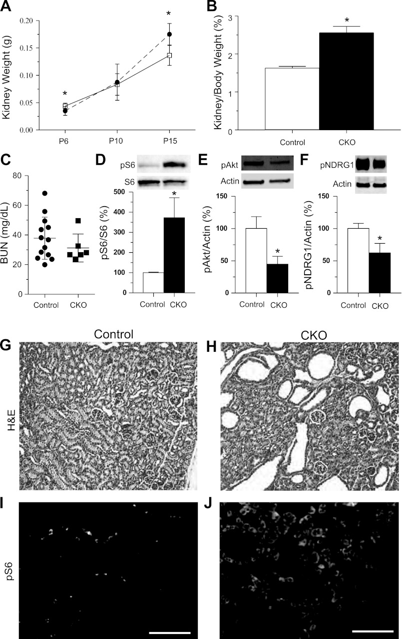
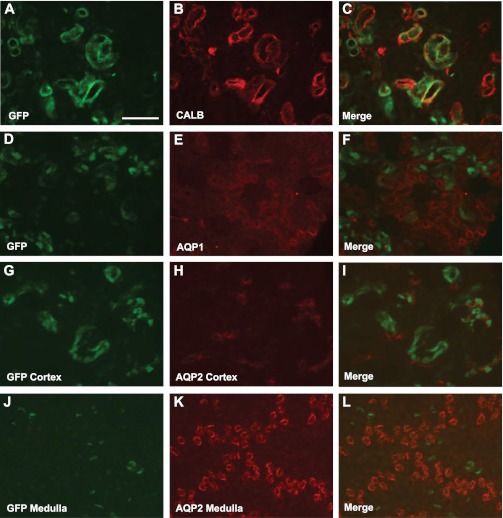
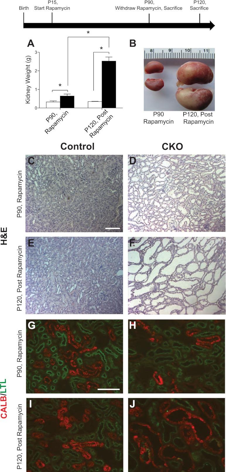
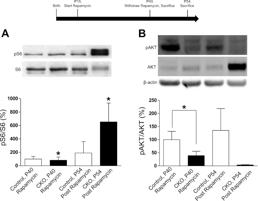
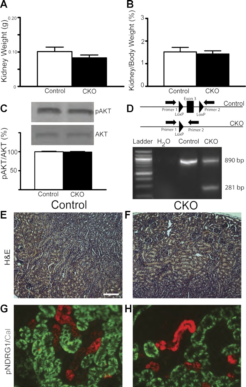
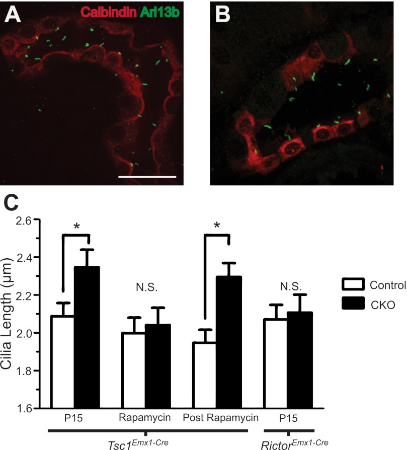
Tuberous sclerosis complex (TSC) is a multiorgan hamartomatous disease caused by loss of function mutations of either the TSC1 or TSC2 genes. Neurological symptoms of TSC predominate in younger patients, but renal pathologies are a serious aspect of the disease in older children and adults. To study TSC pathogenesis in the kidney, we inactivated the mouse Tsc1 gene in the distal convoluted tubules (DCT). At young ages, Tsc1 conditional knockout (CKO) mice have enlarged kidneys and mild cystogenesis with increased mammalian target of rapamycin complex (mTORC)1 but decreased mTORC2 signaling. Treatment with the mTORC1 inhibitor rapamycin reduces kidney size and cystogenesis. Rapamycin withdrawal led to massive cystogenesis involving both distal as well as proximal tubules. To assess the contribution of decreased mTORC2 signaling in kidney pathogenesis, we also generated Rictor CKO mice. These animals did not have any detectable kidney pathology. Finally, we examined primary cilia in the DCT. Cilia were longer in Tsc1 CKO mice, and rapamycin treatment returned cilia length to normal. Rictor CKO mice had normal cilia in the DCT. Overall, our findings suggest that loss of the Tsc1 gene in the DCT is sufficient for renal cystogenesis. This cytogenesis appears to be mTORC1 but not mTORC2 dependent. Intriguingly, the mechanism may be cell autonomous as well as non-cell autonomous and possibly involves the length and function of primary cilia.


Figures

Fig. 1.
Fig. 1.
Postnatal day (P)15 Tsc1 conditional knockout (CKO) mice have large kidneys, renal cysts and increased mammalian target of rapamycin complex 1 (mTORC1) signaling. A: kidney weights from control (solid line) or Tsc1 CKO mice (dashed line) at P6 to P15; n ≥ 3 for each group. *P < 0.03. B: kidney weight normalized to body weight from P15 control and Tsc1 CKO mice. (control n = 9; CKO n = 3). *P < 0.03. C: no differences in blood urea nitrogen (BUN) levels from control and Tsc1 CKO mice. *P > 0.05, Student's t-test (control n = 13; CKO n = 6). D: immunoblotting for phospho-S6 reveals increased mTORC1 activity. Blots were stripped and reprobed for total S6 levels. E: immunoblotting for phospho-Akt (Ser473) reveals decreased mTORC2 signaling. F: decreased phospho-NDRG1 (Thr346) further reveals decreased mTORC2 signaling in P15 Tsc1 CKO kidney compared with littermate controls. Data were analyzed with Student's t-test. *P < 0.001 for phospho-S6, P < 0.05 for phospho-Akt and *P < 0.005 for phospho-NDRG1. All graphs are plotted as means ± SD, control extract expression levels were set to 100%. G and H: hematoxylin and eosin (H&E) staining of kidney sections from P15 control and Tsc1 CKO mice. Moderate cystic dilations are seen in kidneys from Tsc1 CKO mice. I and J: immunofluorescence for phospho-S6 in control and Tsc1 CKO mice. Phospho-S6 is diffusely expressed in the majority of the Tsc1 CKO kidney but found only in isolated cells in kidney from control littermates. Scale bars = 100 μm.
Fig. 2.
Fig. 2.
Emx1-Cre mediates gene recombination in the distal convoluted tubule. AC: GFP expression from Cre recombinase fate mapping (A) and calbindin (CALB), a marker of the distal convoluted tubules (DCT; B), colocalize in the cortex of the kidney, merged image (C). Neither AQP1, a marker of the proximal tubule, nor aquaporin 2 (AQP2), a marker of collecting ducts, colocalize in the cortex with GFP (DF and GI, respectively). Only faint reporter GFP was observed in the medulla, and this signal did not colocalize with AQP2 (JL). Scale bar = 100 μm.
Fig. 3.
Fig. 3.
Rapamycin treatment partially inhibits cyst formation in Tsc1 CKO mice and upon withdrawal causes giant cystic kidneys. Two groups of mice were treated with rapamycin from P15 to P90. At P90, kidneys from one group of animals euthanized on rapamycin were analyzed. For the second group of animals, rapamycin treatment was discontinued at P90 and mice were euthanized 30 days postrapamycin treatment at P120. A: rapamycin treatment reduces cystogenesis in Tsc1 CKO kidneys. After rapamycin withdrawal Tsc1 CKO kidneys become much larger than those from control littermates (n ≥ 3 for each group of control and Tsc1 CKO mice). B: gross images of representative Tsc1 CKO kidneys on rapamycin at P90 (left) or 30 days postrapamycin treatment at P120 (right). CF: H&E staining of paraffin sections from Tsc1 CKO and control littermate kidneys on or off of rapamycin. Rapamycin-treated Tsc1 CKO mice have slightly dilated tubules at P90 (D) compared with controls (C). At P120, 30 days postrapamycin treatment, kidneys from Tsc1 CKO show large cystic dilations of all tubules (F) compared with littermate controls (E). GJ: immunofluorescence for LTL [proximal convoluted tubule (PCT) marker, green] and the calbindin (DCT marker, red). Dilations of both the PCT and DCT are observed while on rapamycin (H) and postrapamycin treatment (J) compared with controls (G and I, respectively). Graphs are plotted with means ± SD. Data were analyzed with Student's t-test. *P < 0.001. Scale bar = 100 μm.
Fig. 4.
Fig. 4.
Rapamycin treatment normalizes mTORC1 but not mTORC2 signaling in Tsc1 CKO kidneys. Two groups of animals were treated with rapamycin from P15 to P40. Kidneys were taken from the first group of euthanized animals at P40 (n = 4 for control mice and n = 3 for CKO). Rapamycin treatment was discontinued in the second group of animals for 14 days, from P40 to P54 and protein extracts made from these kidneys at P54 (n = 3 for control and n = 2 for CKO). A: mTORC1 signaling, as shown by S6 protein phosphorylation, in Tsc1 CKO kidneys was decreased to that seen in control animals with rapamycin treatment. Postrapamycin phospho-S6 levels were greatly increased. *P < 0.05. B: mTORC2 signaling, as shown by Akt phosphorylation at Serine473, in P40 Tsc1 CKO kidneys was significantly decreased while on rapamycin. *P = 0.039. While trending towards significance, CKO extracts postrapamycin treatment did not have statistically significant decreases in phospho-Akt (Serine473); P = 0.12.
Fig. 5.
Fig. 5.
Rictor CKO kidneys at P15 do not develop cysts. A and B: kidneys from P15 Rictor CKO mice did not show alterations in kidney weight nor in kidney-to-body weight ratios compared with control littermates (control n = 15; CKO n = 7). C: no changes in AKT phosphorylation at Serine 473 were detected through western blot analyses of protein extracts from P9-P15 Rictor CKO mice. D: PCR reveals recombination of exon three of the Rictor floxed allele in the kidney of Rictor CKO mice. E and F: H&E staining do not show any appreciable kidney abnormalities. G and H: coimmunofluorescence for pNDRG1, a marker of mTORC2 activity, and calbindin, a marker of the DCT, revealed undetectable levels of mTORC2 activity in the DCT in both control and CKO animals.
Fig. 6.
Fig. 6.
Primary cilia are longer in Tsc1-deficient but not Rictor-deficient tubules. A and B: representative photomicrographs of cilia in the DCT of Tsc1 CKO mice on rapamycin at P90 (A) or 30 days after rapamycin withdrawal at P120 (B). Coimmunofluorescence for Arl13B (a marker of primary cilia) and calbindin (marker of the DCT). C: cilia length in untreated mice at P15, during rapamycin treatment at P90 and 30 days postrapamycin withdrawal at P120. Primary cilia in the DCT of kidneys from Tsc1 CKO mice were longer 30 days after rapamycin cessation. No changes in cilia length were noted between control mice and Rictor CKO mice. *P < 0.05. Scale bar = 25 μm. N.S, not statistically significant; n = 3 for each group of control, Tsc1 CKO and RictorEmx1−Cre CKO mice.

References

    1. Badano JL, Mitsuma N, Beales PL, Katsanis N. The ciliopathies: an emerging class of human genetic disorders. Annu Rev Genomics Hum Genet 7: 125– 148, 2006 - PubMed
    1. Briata P, Di Blas E, Gulisano M, Mallamaci A, Iannone R, Boncinelli E, Corte G. EMX1 homeoprotein is expressed in cell nuclei of the developing cerebral cortex and in the axons of the olfactory sensory neurons. Mech Dev 57: 169– 180, 1996 - PubMed
    1. Carson RP, Van Nielen DL, Winzenburger PA, Ess KC. Neuronal and glia abnormalities in Tsc1-deficient forebrain and partial rescue by rapamycin. Neurobiol Dis 45: 369– 380, 2012 - PMC - PubMed
    1. Consortium of European Chromosome 16 Tuberous Sclerosis Identification and characterization of the tuberous sclerosis gene on chromosome 16. Cell 75: 1305– 1315, 1993 - PubMed
    1. Crino PB, Aronica E, Baltuch G, Nathanson KL. Biallelic TSC gene inactivation in tuberous sclerosis complex. Neurology 74: 1716– 1723, 2010 - PMC - PubMed

Publication types

MeSH terms

LinkOut - more resources