. 2012 Dec;61(12):2239-49.
doi: 10.1007/s00262-012-1290-9. Epub 2012 Jun 7.

Cellular and cytokine-dependent immunosuppressive mechanisms of grm1-transgenic murine melanoma

Affiliations

Cellular and cytokine-dependent immunosuppressive mechanisms of grm1-transgenic murine melanoma

Miriam Alb et al. Cancer Immunol Immunother. 2012 Dec.

Abstract

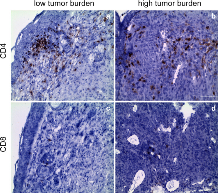
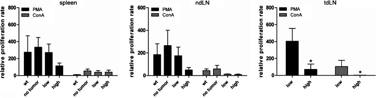
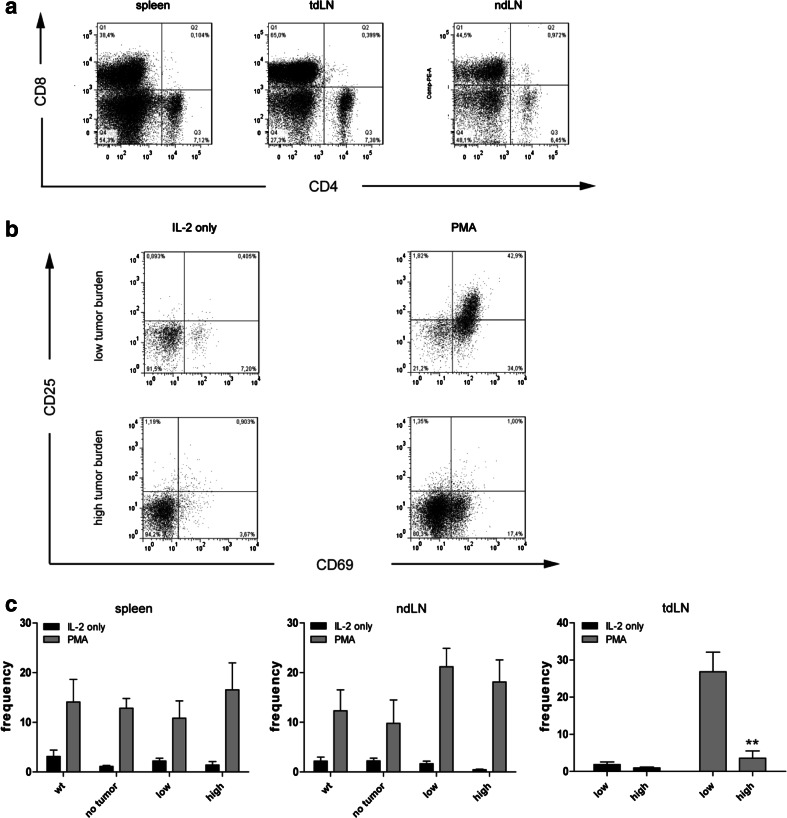
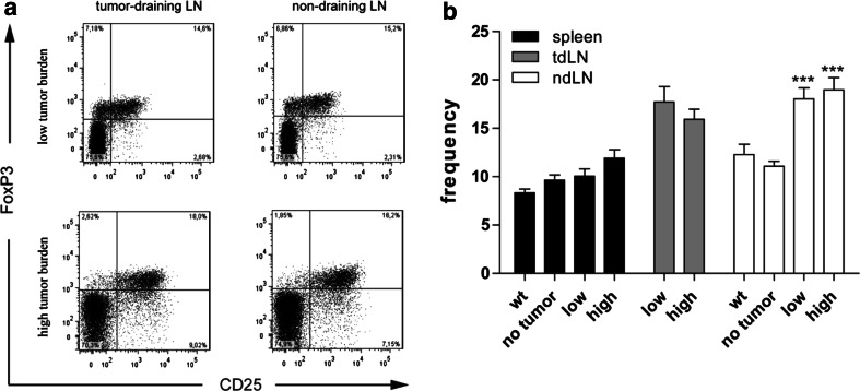
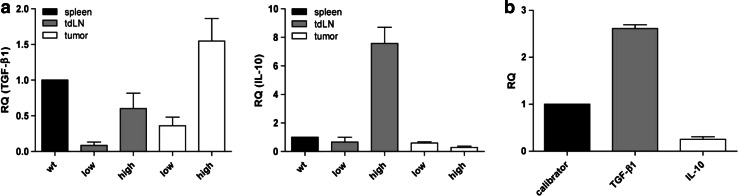
Grm1-transgenic mice spontaneously develop cutaneous melanoma. This model allowed us to scrutinize the generic immune responses over the course of melanoma development. To this end, lymphocytes obtained from spleens, unrelated lymph nodes and tumor-draining lymph nodes of mice with no evidence of disease, and low or high tumor burden were analyzed ex vivo and in vitro. Thereby, we could demonstrate an increase in the number of activated CD4(+) and CD8(+) lymphocytes in the respective organs with increasing tumor burden. However, mainly CD4(+) T cells, which could constitute both T helper as well as immunosuppressive regulatory T cells, but not CD8(+) T cells, expressed activation markers upon in vitro stimulation when obtained from tumor-bearing mice. Interestingly, these cells from tumor-burdened animals were also functionally hampered in their proliferative response even when subjected to strong in vitro stimulation. Further analyses revealed that the increased frequency of regulatory T cells in tumor-bearing mice is an early event present in all lymphoid organs. Additionally, expression of the immunosuppressive cytokines TGF-β1 and IL-10 became more evident with increased tumor burden. Notably, TGF-β1 is strongly expressed in both the tumor and the tumor-draining lymph node, whereas IL-10 expression is more pronounced in the lymph node, suggesting a more complex regulation of IL-10. Thus, similar to the situation in melanoma patients, both cytokines as well as cellular immune escape mechanisms seem to contribute to the observed immunosuppressed state of tumor-bearing grm1-transgenic mice, suggesting that this model is suitable for preclinical testing of immunomodulatory therapeutics.


Figures

Fig. 1
Fig. 1
Tumor-infiltrating lymphocytes (TIL) in LLA-TG-3 tumors. a, b Showing CD4+ T cells in LLA-TG-3 tumors (red staining); c, d showing CD8+ T cells in LLA-TG-3 tumors (red staining). Nuclei were stained using hematoxylin. Scale bar: 100 μm. a, c Low tumor burden; b, d high tumor burden
Fig. 2
Fig. 2
Gating strategy; CD4+ and CD8+ T-cell subsets ex vivo. a Gating strategy used for all flow cytometry data shown; bd frequency of activated CD4+ T-cell subsets in spleen and lymph nodes of wild-type (wt) and LLA-TG-3 mice; eg frequency of activated CD8+ T-cell subsets in spleen and lymph nodes of wt and LLA-TG-3 mice; n = 14 for wt mice; n = 10 for no tumor and low tumor burden LLA-TG-3 mice; n = 13 for high tumor burden LLA-TG-3 mice. Depicted are mean values + SEM. *p < 0.05; **p < 0.005
Fig. 3
Fig. 3
Proliferative response of lymphocytes upon mitogen stimulation. The proliferation rate of splenocytes and lymph node cells was analyzed using a 3H-thymidine incorporation assay after incubation for 4 days in medium supplemented with IL-2 (500 IU/ml) and PMA (200 nM) or ConA (5 μg/ml); n = 11 for wt mice; n = 9 for no tumor LLA-TG-3, and n = 10 for low and high tumor burden LLA-TG-3 mice. The proliferation rate was calculated relative to the proliferation rate in medium supplemented with IL-2 (500 IU/ml) only. Depicted are mean values + SEM. *p < 0.05
Fig. 4
Fig. 4
Frequency of CD4+ and CD8+ T cells upon IL-2 and PMA stimulation. a Representative dot plots of CD4 and CD8 stainings in spleen, tumor-draining (tdLN), and non-draining (ndLN) lymph nodes of a high tumor burden LLA-TG-3 mouse upon IL-2/PMA stimulation; b representative flow cytometry analysis of gated CD8+ T cells in tumor-draining LN of low and high tumor burden LLA-TG-3 mice upon stimulation with IL-2 (500 IU/ml) or IL-2 and PMA (200 nM). Numbers in top right quadrants indicate CD69+CD25+ cells; c the frequency of CD69+/CD25+ CD8+ T cells in spleen, tdLN, and ndLN of wt and LLA-TG-3 mice was analyzed as in a; n = 10 for wt mice; n = 6 for no tumor and low tumor burden LLA-TG-3 mice; n = 8 for high tumor burden LLA-TG-3 mice. Depicted are mean values + SEM. **p < 0.005
Fig. 5
Fig. 5
Frequency of regulatory T cells ex vivo (Tregs; CD4+CD25+FoxP3+). a Representative flow cytometry analysis of gated CD4+ T cells in tdLN and ndLN of tumor-bearing LLA-TG-3 mice. Numbers in top right quadrants indicate CD25+FoxP3+ cells; b the frequency of Tregs was analyzed as in a in spleens and LN of wt and LLA-TG-3 mice ex vivo; n = 13 for wt mice; n = 10 for no tumor and low tumor burden LLA-TG-3 mice; n = 11 for high tumor burden LLA-TG-3 mice. Depicted are mean values + SEM. ***p < 0.0001
Fig. 6
Fig. 6
Relative mRNA expression of TGF-β1 and IL-10. a The mRNA levels of TGF-β1 (left) and IL-10 (right) relative to wt splenocytes were analyzed using qRT-PCR and cDNA from splenocytes, lymph node cells, and tumor samples of wt and LLA-TG-3 mice; n = 1 for wt mice; n = 3 for low tumor burden LLA-TG-3 mice; n = 4 for high tumor burden LLA-TG-3 mice; b the mRNA levels of TGF-β1 and IL-10 relative to the immortalized melanocyte cell line B10.BR (calibrator) were analyzed using qRT-PCR and cDNA from the two LLA-TG-3 tumor cell lines Nu2 and 4046T. Depicted are mean values + SEM

References

    1. Dunn GP, Old LJ, Schreiber RD. The three Es of cancer immunoediting. Annu Rev Immunol. 2004;22:329–360. doi: 10.1146/annurev.immunol.22.012703.104803. - DOI - PubMed
    1. Ramirez-Montagut T, Turk MJ, Wolchok JD, Guevara-Patino JA, Houghton AN. Immunity to melanoma: unraveling the relation of tumor immunity and autoimmunity. Oncogene. 2003;22:3180–3187. doi: 10.1038/sj.onc.1206462. - DOI - PubMed
    1. Andersen MH, Pedersen LO, Capeller B, Brocker EB, Becker JC, thor Straten P. Spontaneous cytotoxic T-cell responses against survivin-derived MHC class I-restricted T-cell epitopes in situ as well as ex vivo in cancer patients. Cancer Res. 2001;61:5964–5968. - PubMed
    1. Zhang L, Conejo-Garcia JR, Katsaros D, et al. Intratumoral T cells, recurrence, and survival in epithelial ovarian cancer. N Engl J Med. 2003;348:203–213. doi: 10.1056/NEJMoa020177. - DOI - PubMed
    1. van Houdt IS, Sluijter BJ, Moesbergen LM, et al. Favorable outcome in clinically stage II melanoma patients is associated with the presence of activated tumor infiltrating T-lymphocytes and preserved MHC class I antigen expression. Int J Cancer. 2008;123:609–615. doi: 10.1002/ijc.23543. - DOI - PubMed

MeSH terms

LinkOut - more resources