Review
. 2012:60:279-99.
doi: 10.1007/978-94-007-4186-7_12.

Adherens junctions in C. elegans embryonic morphogenesis

Affiliations
Review

Adherens junctions in C. elegans embryonic morphogenesis

Stephen T Armenti et al. Subcell Biochem. 2012.

Abstract

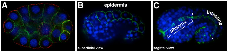
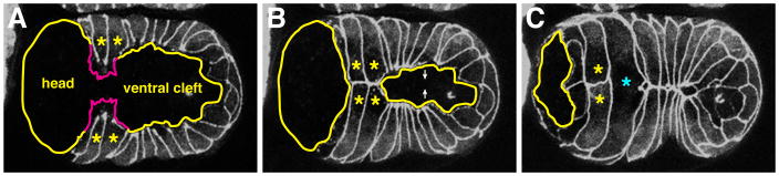
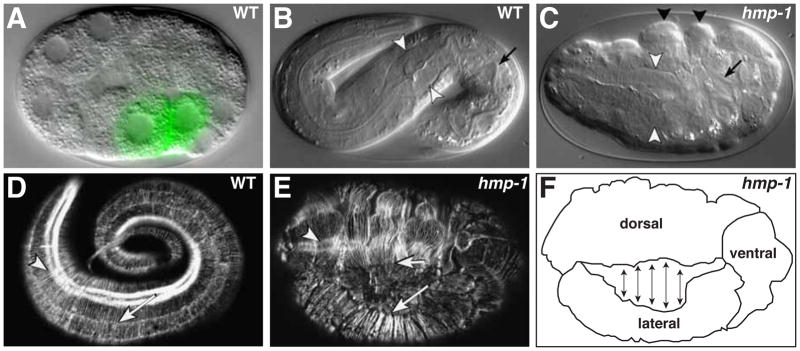
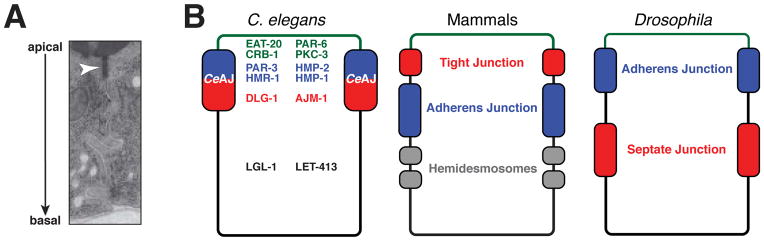
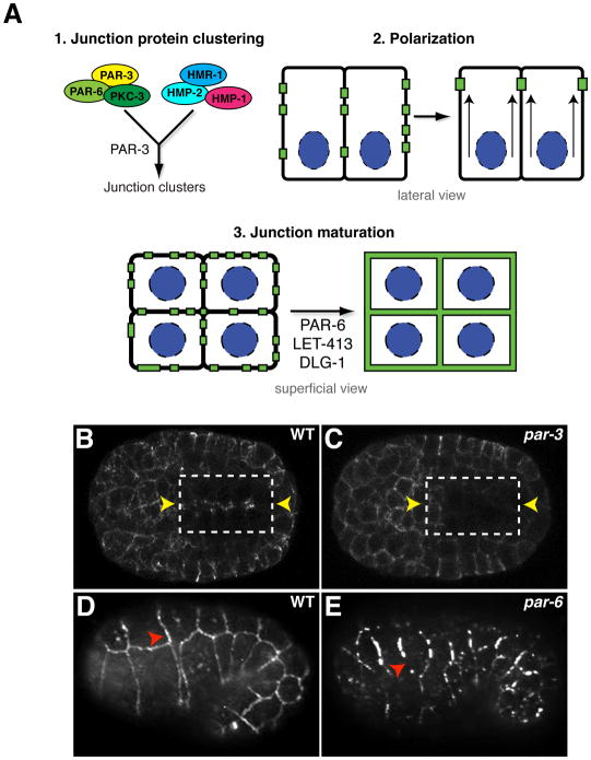
Caenorhabditis elegans provides a simplified, in vivo model system in which to study adherens junctions (AJs) and their role in morphogenesis. The core AJ components-HMR-1/E-cadherin, HMP-2/β-catenin and HMP-1/α-catenin-were initially identified through genetic screens for mutants with body axis elongation defects. In early embryos, AJ proteins are found at sites of contact between blastomeres, and in epithelial cells AJ proteins localize to the multifaceted apical junction (CeAJ)-a single structure that combines the adhesive and barrier functions of vertebrate adherens and tight junctions. The apically localized polarity proteins PAR-3 and PAR-6 mediate formation and maturation of junctions, while the basolaterally localized regulator LET-413/Scribble ensures that junctions remain apically positioned. AJs promote robust adhesion between epithelial cells and provide mechanical resistance for the physical strains of morphogenesis. However, in contrast to vertebrates, C. elegans AJ proteins are not essential for general cell adhesion or for epithelial cell polarization. A combination of conserved and novel proteins localizes to the CeAJ and works together with AJ proteins to mediate adhesion.


Figures

Figure 1
Figure 1. HMR-1/E-cadherin localization in blastomeres and epithelial cells
All panels show immunostained embryos; DNA is stained blue. (A) HMR-1 (green) in early embryos localizes to sites of contact between blastomeres (a 26-cell stage embryo is shown). Contact-free surfaces are marked with PAR-3 (red). (B–C) After epithelial cells begin to form during mid-embryogenesis, HMR-1 localizes to adherens junctions. (B) Superficial view showing HMR-1 at junctions between epidermal cells. (C) Sagittal view of central region of embryo showing HMR-1 at junctions in pharyngeal and intestinal epithelial cells, which form a tube comprising the digestive tract; region of intestinal cells is bounded by arrowheads.
Figure 2
Figure 2. Ventral Enclosure
(A) Embryo near the start of ventral enclosure expressing DLG-1-GFP to mark junctions, ventral view. Leading cells are marked by asterisks and filopodia are depicted by magenta tracings. The ventral cleft and future head region are indicated. (B) Middle of ventral enclosure. Leading cells have generated nascent contacts with contralateral cells. The ventral cleft is closing as pocket cells begin to come together. (C) End of ventral enclosure. The ventral cleft has closed and the posterior pair of leading cells has fused (cyan asterisk), abolishing the initial contact. Images reprinted from Chisholm and Hardin, 2005.
Figure 3
Figure 3. Gastrulation and epidermal morphogenesis
(A) Early embryo at the onset of gastrulation. The E daughters (labeled green by end-1::GFP transgene) have flattened their apical surfaces and are beginning to ingress into the embryo. (B) Fully elongated wildtype embryo. In B and C, white arrowheads indicate pharyngeal bulb and arrow marks the intestinal lumen. (C) hmp-1 mutant embryo displaying the Humpback (Hmp) phenotype, with characteristic dorsal epidermal bulges (black arrowheads). Note shortened pharynx and intestine (arrow) due to elongation failure. (D) Fully elongated wildtype embryo stained with phalloidin to visualize F-actin. Arrow indicates parallel bundles of circumferential actin filaments, arrowhead indicates actin filaments in underlying muscle tissue. (E) Phalloidin-stained hmp-1 mutant embryo. Circumferential actin bundles between dorsal (short arrow) and lateral (long arrow) epidermal cells have detached. Arrowhead indicates intact underlying muscle actin filaments. (F) Schematic diagram of embryo in E showing points of separation between dorsal and lateral epidermal cells. Images in panels B-E are reprinted from Costa et al., 1998; Panel F was redrawn from a similar panel in Costa et al., 1998.
Figure 4
Figure 4. The C. elegans apical junction and analogous junctions in mammals and Drosophila
(A) Transmission electron micrograph of intestinal epithelial cells showing the C. elegans apical junction (CeAJ, arrowhead) as a single electron-dense region. (B) Schematic diagram of epithelial domains and junction structures in C. elegans, mammals and Drosophila. Major polarity and junction proteins and their localization pattern in mature C. elegans epithelia are depicted. Functionally analogous regions in mammals and Drosophila are shown in common colors. Panel A reprinted from Muller and Bossinger, 2003.
Figure 5
Figure 5. Adherens junction formation
(A) Junction formation involves distinct steps: (1) PAR-3 is required for clustering adherens junction proteins early during polarization. PAR proteins and adherens junctions proteins are all present in puncta. (2) Epithelial cells polarize and junction clusters are recruited apically to the site of future junction formation. (3) Apically localized puncta mature into belt-like junctions through the function of PAR-6, LET-413, and DLG-1. (B, C) Polarization of junction clusters in WT (B) and embryos lacking both maternal and zygotic PAR-3 (C). HMR-1-GFP is shown. In each panel, the box represents the polarizing intestine. Left and right rows of intestinal cells show mirror symmetry, and the future apical surface between them is indicated by yellow arrowheads. (D,E) Junction maturation in wildtype (D) and embryos lacking both maternal and zygotic PAR-6 (E). Red arrowheads indicate continuous junctions in wildtype (D) and fragmented junctions in par-6 mutants. Junctions are stained with DLG-1, and the epidermis is shown. Panels B-E are reprinted from Achilleos et al., 2010.

References

    1. Aceto D, Beers M, Kemphues KJ. Interaction of PAR-6 with CDC-42 is required for maintenance but not establishment of PAR asymmetry in C. elegans. Dev Biol. 2006;299:386–397. - PMC - PubMed
    1. Achilleos A, Wehman AM, Nance J. PAR-3 mediates the initial clustering and apical localization of junction and polarity proteins during C. elegans intestinal epithelial cell polarization. Development. 2010;137:1833–1842. - PMC - PubMed
    1. Adams CL, Chen YT, Smith SJ, Nelson WJ. Mechanisms of epithelial cell-cell adhesion and cell compaction revealed by high-resolution tracking of E-cadherin-green fluorescent protein. J Cell Biol. 1998;142:1105–1119. - PMC - PubMed
    1. Anastasiadis PZ. p120-ctn: A nexus for contextual signaling via Rho GTPases. Biochim Biophys Acta. 2007;1773:34–46. - PubMed
    1. Aono S, Legouis R, Hoose WA, Kemphues KJ. PAR-3 is required for epithelial cell polarity in the distal spermatheca of C. elegans. Development. 2004;131:2865–2874. - PubMed

MeSH terms

Substances

LinkOut - more resources