. 2012 Aug;27(8):2341-51.
doi: 10.1093/humrep/des196. Epub 2012 Jun 6.

Lesion kinetics in a non-human primate model of endometriosis

Affiliations

Lesion kinetics in a non-human primate model of endometriosis

P Harirchian et al. Hum Reprod. 2012 Aug.

Abstract

Background: Endometriosis is a common cause of pelvic pain and infertility in women of reproductive age. It is characterized by the presence of endometrial tissue outside the normal location, predominantly in the pelvic peritoneum causing severe abdominal pain. However, the severity of the symptoms of endometriosis does not always correlate with the anatomic severity of the disease. This lack of correlation may be due to morphological lesion variation during disease progression. This study examined lesion kinetics in a non-human primate model of endometriosis to better understand lesion dynamics.

Methods: Endometriosis was experimentally induced in nine normal cycling female adult olive baboons (Papio anubis) by i.p. inoculation of autologous menstrual endometrium on Day 2 of menses for two consecutive menstrual cycles. Diagnostic laparoscopies were performed between Day 8-12 post-ovulation at 1, 3, 6, 9 and 12 months, followed by a necropsy at 15 months, after the second inoculation. In two animals, lesions were excised/ablated at 6 months and they were monitored for lesion recurrence and morphological changes by serial laparoscopy. Furthermore, five control animals underwent surgeries conducted at the same time points but without inoculation.

Results: A total of 542 endometriotic lesions were observed. The location, macroscopic (different colours) and microscopic appearance confirmed distinct endometriosis pathology in line with human disease. The majority of the lesions found 1 month after tissue inoculation were red lesions, which frequently changed colour during the disease progression. In contrast, blue lesions remained consistently blue while white lesions were evident at the later stages of the disease process and often regressed. There were significantly lower numbers of powder burn, blister and multicoloured lesions observed per animal in comparison to black and blue lesions (P-value≤0.05). New lesions were continually arising and persisted up to 15 months post-inoculation. Lesions reoccurred as early as 3 months after removal and 69% of lesions excised/ablated had reoccurred 9 months later. Interestingly, endometriotic lesions were also found in the non-inoculated animals, starting at the 6-month time point following multiple surgeries.

Conclusions: Documentation of lesion turnover in baboons indicated that lesions changed their colour from red to white over time. Different lesion types underwent metamorphosis at different rates. A classification of lesions based on morphological appearance may help disease prognosis and examination of the effect of the lesion on disease symptoms, and provide new opportunities for targeted therapies in order to prevent or treat endometriosis. Surgical removal of endometriotic lesions resulted in a high incidence of recurrence. Spontaneous endometriosis developed in control baboons in the absence of inoculation suggesting that repetitive surgical procedures alone can induce the spontaneous evolution of the chronic disease. Although lesion excision/ablation may have short-term benefits (e.g. prior to an IVF cycle in subfertile women), for long-term relief of symptoms perhaps medical therapy is more effective than surgical therapy.


Figures

Figure 1
Figure 1
Experimental model of endometriosis in baboons, design and timeline. (A) Nine baboons were inoculated with autologous menstrual tissue at two consecutive menses. (B) Two baboons with induced endometriosis had all lesions excised/ablated at 6-month laparoscopy. (C) Five animals did not undergo inoculation, but were subjected to the same surgical procedures as group A. All animals underwent diagnostic laparoscopies accompanied with an endometrectomy between Days 8–12 post-ovulation at 1, 3, 6, 9 and 12 months followed by necropsy at 15 months after the second inoculation.
Figure 2
Figure 2
Endometriotic lesion location and morphology in an experimental model of endometriosis in baboons. Nine baboons were experimentally induced with endometriosis by i.p. inoculation with autologous menstrual endometrium, as described in Materials and Methods. (A) Common locations of baboon endometriotic lesions in the abdomen were the perimetrium, the bladder and the peritoneum at the Pouch of Douglas. (B) Gomori trichrome staining revealed distinct endometrial glands and stroma. Haemorrhaging was observed in the red lesions (arrow), while more connective tissue was present in the white lesions (arrow). Scale bar: 100 μm.
Figure 3
Figure 3
Number of new lesions and their persistence over time in an experimental model of endometriosis in baboons. Nine baboons underwent diagnostic laparoscopies at 1, 3, 6, 9 and 12 months as well as a necropsy at 15 months following two i.p. inoculations with autologous menstrual endometrium. Each lesion was recorded upon initial sighting and the site was specifically assessed at subsequent laparoscopies. (A) Mean number of new lesions per inoculated animal at each laparoscopy and 15-month necropsy. (B) The mean number of lesions first seen at each laparoscopy in the inoculated animals that are still present at 15-month necropsy. Columns represent the mean number of lesions per animal (n= 9). Error bars depict SD. ns, not significant (one-way ANOVA Tukey's multiple comparison post hoc test).
Figure 4
Figure 4
Number of endometriotic lesions characterized by different colours in an experimental model of endometriosis in baboons. Each lesion was recorded upon initial sighting and tracked at each subsequent laparoscopy where colours were recorded. (A) There were significantly lower numbers of powder burn, blister and multicoloured lesions observed per animal in comparison to black and blue lesions (*P-value ≤ 0.05, one-way ANOVA Tukey's multiple comparison post hoc test). Columns represent the mean number of lesions per animal (n= 9). Error bars represent the SD of the mean. (B) Frequency of lesions (in % of total number of lesions per time point) characterized by different colours in the nine baboons at different time points post-inoculation.
Figure 5
Figure 5
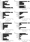
Turnover of endometriotic lesions characterized by different colours in an experimental model of endometriosis in baboons. At each surgery the locations of previously identified endometriotic lesions were analyzed and the evolution of each lesion tracked. When the site of the lesion was not accessible, the lesion was categorized as not found/not visible. When the site of the lesion was accessible and no lesion was observed, the lesion was categorized as regressed (not present). (A, C, E and G) Show the number of red, black, blue and white lesions at each laparoscopy. (B, D, F and H) Demonstrate the evolution of red, black, blue and white lesions at the subsequent laparoscopy. The bars correspond to the total number of lesions per time point or lesion type, respectively.
Figure 6
Figure 6
Recurrence of excised/ablated endometriotic lesions in an experimental model of endometriosis in baboons. In two animals that were inoculated with autologous menstrual tissue, lesions were excised or ablated 6 months after the second inoculation. Laparoscopies were performed at 9 and 12 months and a necropsy at 15 months to control for lesion recurrence. (A) Of the removed lesions at 6 months post-inoculation, 3 of 16 returned 3 months after excision, 7 of 26 returned 6 months after excision and at 15-month necropsy 18 of 26 (69%) lesions had returned. (B) Excision and ablation were used interchangeably as determined by the surgeon. Fifteen of 20 (75%) excised lesions returned by 15-month necropsy while three of six (50%) ablated lesions returned by necropsy.
Figure 7
Figure 7
Turnover of reoccurring excised/ablated endometriotic lesions in an experimental model of endometriosis in baboons. Excision/ablation of all visible endometriotic lesions was performed in two baboons 6 months after experimental induction of endometriosis, as described in Materials and Methods (Fig. 1). Laparoscopies were conducted at 9 and 12 months and a necropsy at 15 months post-inoculation to determine the incidence of lesion recurrence and macroscopic changes within the peritoneal cavity. When the site of the lesion was not accessible, the lesion was categorized as not found/not visible. When the site of the lesion was accessible and no lesion was observed, the lesion was categorized as regressed (not present). Black and blue bars represent the status of excised/ablated black and blue endometriotic lesions after recurrence. (A) The reoccurring excised/ablated black and blue lesions at subsequent laparoscopy. Each bar represents the number of each type of lesion manifested after reoccurrence. (B) The reoccurring excised/ablated black and blue lesions at 15-month necropsy. Each bar represents the number of each type of lesion manifested at necropsy.
Figure 8
Figure 8
Control baboons not inoculated with menstrual tissue. Five animals did not undergo inoculation, but were subjected to the same schedule of surgeries as the experimental groups to control for the effects of the surgeries themselves. Twenty-seven endometriotic lesions were observed in the five animals. (A) Each bar represents the total number of endometriotic lesions found per animal. (B) Each bar represents the number of newly detected lesions at the corresponding time point. (C) Each bar represents the total number of each lesion type observed.

References

    1. Abbott JA, Hawe J, Clayton RD, Garry R. The effects and effectiveness of laparoscopic excision of endometriosis: a prospective study with 2–5 year follow-up. Hum Reprod. 2003;18:1922–1927. - PubMed
    1. American Fertility Society. Revised American Fertility Society classification of endometriosis 1985. Fertil Steril. 1985;43:351–352. - PubMed
    1. American Fertility Society. Revised American Society for Reproductive Medicine classification of endometriosis: 1996. Fertil Steril. 1997;67:817–821. - PubMed
    1. Braundmeier AG, Fazleabas AT. The non-human primate model of endometriosis: research and implications for fecundity. Mol Hum Reprod. 2009;15:577–586. - PMC - PubMed
    1. Brosens I. Endometriosis rediscovered? Hum Reprod. 2004;19:1679–1680. author reply 1680–1671. - PubMed

Publication types