. 2012 Oct;64(10):3334-43.
doi: 10.1002/art.34556.

The mesenchymal stem cell marker CD248 (endosialin) is a negative regulator of bone formation in mice

Affiliations

The mesenchymal stem cell marker CD248 (endosialin) is a negative regulator of bone formation in mice

Amy J Naylor et al. Arthritis Rheum. 2012 Oct.

Abstract

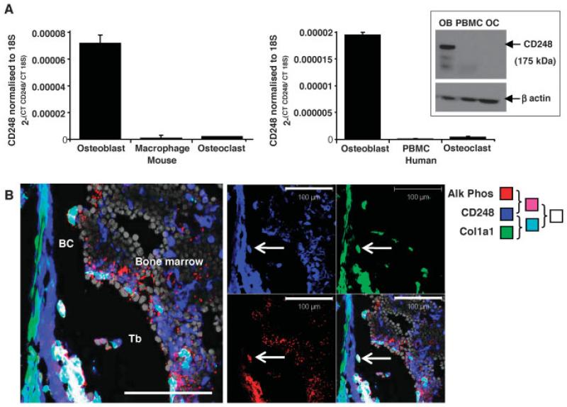
Objective: CD248 (tumor endothelial marker 1/endosialin) is found on stromal cells and is highly expressed during malignancy and inflammation. Studies have shown a reduction in inflammatory arthritis in CD248-knockout (CD248(-/-) ) mice. The aim of the present study was to investigate the functional effect of genetic deletion of CD248 on bone mass.

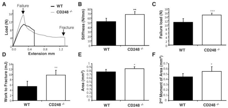
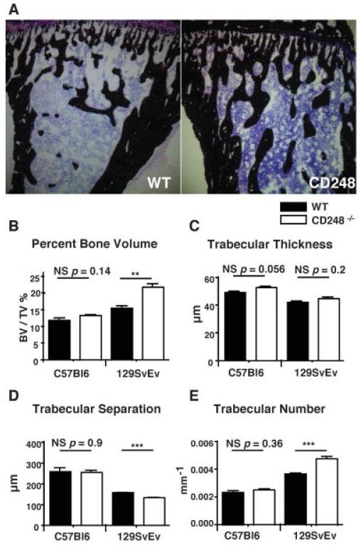
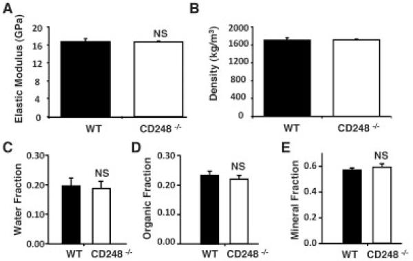
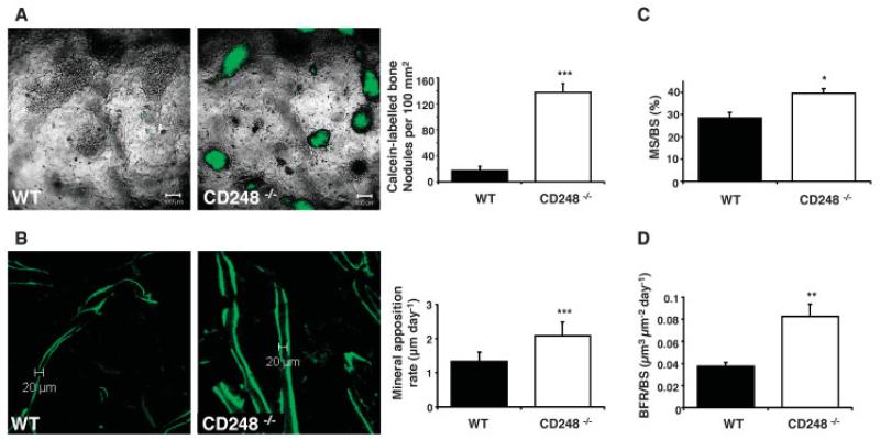
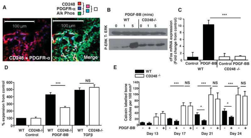
Methods: Western blotting, polymerase chain reaction, and immunofluorescence were used to investigate the expression of CD248 in humans and mice. Micro-computed tomography and the 3-point bending test were used to measure bone parameters and mechanical properties of the tibiae of 10-week-old wild-type (WT) or CD248(-/-) mice. Human and mouse primary osteoblasts were cultured in medium containing 10 mM β-glycerophosphate and 50 μg/ml ascorbic acid to induce mineralization, and then treated with platelet-derived growth factor BB (PDGF-BB). The mineral apposition rate in vivo was calculated by identifying newly formed bone via calcein labeling.

Results: Expression of CD248 was seen in human and mouse osteoblasts, but not osteoclasts. CD248(-/-) mouse tibiae had higher bone mass and superior mechanical properties (increased load required to cause fracture) compared to WT mice. Primary osteoblasts from CD248(-/-) mice induced increased mineralization in vitro and produced increased bone over 7 days in vivo. There was no decrease in bone mineralization and no increase in proliferation of osteoblasts in response to stimulation with PDGF-BB, which could be attributed to a defect in PDGF signal transduction in the CD248(-/-) mice.

Conclusion: There is an unmet clinical need to address rheumatoid arthritis-associated bone loss. Genetic deletion of CD248 in mice results in high bone mass due to increased osteoblast-mediated bone formation, suggesting that targeting CD248 in rheumatoid arthritis may have the effect of increasing bone mass in addition to the previously reported effect of reducing inflammation.


Figures

Figure 1
Figure 1. CD248 is expressed in human and mouse osteoblasts, but not osteoclasts.
A, Real-time polymerase chain reaction was used to analyze the expression of CD248 in primary cell lysates of osteoblasts, macrophages, and osteoclasts from wild-type mice (left) or osteoblasts, peripheral blood mononuclear cells (PBMCs), and osteoclasts from human subjects (right). Bars show the mean ± SD of 3 samples per group. Inset, CD248 expression was also assessed by Western blotting of human cell lysates of osteoblasts (OB), PBMCs, and osteoclasts (OC), using anti-human CD248 (recognizing the band at 175 kd) with anti–β-actin as control (recognizing the band at 47 kd). B, Tibiae from newborn mice were assessed by confocal microscopy for CD248 (blue), Col1a1 (green fluorescent protein under the control of the Col1a1 promoter; green), and alkaline phosphatase (Alk Phos) enzymatic activity (red); nuclei were stained in grey. The merged image in the left panel shows the colocalization of Col1a1 and CD248 (cyan), colocalization of CD248 and alkaline phosphatase (purple), and colocalization of all 3 markers (white). Bar (left panel) = 100 μm. Arrows indicate examples of cells expressing all 3 markers. BC = bone collar; Tb = trabecular bone.
Figure 2
Figure 2. CD248-deficient mice have thicker, stiffer bones that are harder to break.
A–D, Tibiae from 10-week-old wild-type (WT) and CD248−/− C57BL/6 mice were tested to the point of destruction by 3-point bending. Results shown are a representative trace of bone extension as load is applied (A) as well as bone stiffness (B), failure load (C), and work required to fracture (D) in each group. E and F, Micro–computed tomography analysis of the tibiae of 10-week-old WT and CD248−/− C57BL/6 mice was performed to measure cortical bone parameters of cortical area (E) and second moment of area (F). Bars show the mean ± SD of 6 samples per group. * = P < 0.05; ** = P < 0.01; *** = P < 0.001 versus WT, by Student’s t-test.
Figure 3
Figure 3. CD248-deficient mice have increased trabecular bone volume.
A, The tibiae of wild-type (WT) and CD248−/− (129SvEv strain) mice were stained with von Kossa’s stain to detect trabecular bone formation (dark brown; counterstained with hematoxylin [purple]). B–E, Micro–computed tomography was performed to measure the trabecular bone structure in WT and CD248−/− mice on the C57BL/6 and 129SvEv genetic backgrounds, assessed as the percentage of bone volume/tissue volume (BV/TV) (B), trabecular thickness (C), trabecular separation (D), and trabecular number (E). Bars show the mean ± SD of 6 samples per group. ** = P < 0.01; *** = P < 0.001, by Student’s t-test. NS = not significant.
Figure 4
Figure 4. CD248-deficient mice have normal bone composition.
The parameters of bone composition included the elastic modulus (A) and bone density (B), measured in wild-type (WT) and CD248−/− mice on the C57BL/6 background, as well as the water (C), organic matter (D), and mineral (E) fractions, determined using ashing in the C57BL/6 and 129SvEv strains of WT and CD248−/− mice. Bars show the mean ± SD of 6 samples per group. Student’s t-test was used to assess differences between the 2 genotypes. NS = not significant.
Figure 5
Figure 5. Increased osteoblast activity is the cause of increased bone formation in CD248-deficient mice.
A, Cultures of osteoblasts from wild-type (WT) and CD248−/− mice were stained with calcein to label mineralized bone nodules (left), and the bone nodules were quantified in the osteoblast cultures from both groups (right). The experiment was repeated twice; representative results are shown. B, Ten-week-old WT and CD248−/− C57BL/6 mice were given 2 injections of calcein 7 days apart to label mineralizing bone surfaces (left). The distance between these labels, measured in the trabecular bone at the epiphysis of tibia sections, was determined as the mineral apposition rate (right). C and D, The percentage of bone surface that was actively mineralized over the course of the 7-day period of the experiment (mineralized surface/bone surface [MS/BS]) (C) and the bone formation rate per bone surface (BFR/BS) per day (D) were determined in each group. All analyses were performed by investigators blinded with regard to genotype. Bars in A, C, and D show the mean ± SD of 3 samples per group. Bars in B show the mean ± SD of 6 samples per group. * = P < 0.05; ** = P < 0.01; *** = P < 0.001 versus WT, by Student’s t-test.
Figure 6
Figure 6. CD248 signals via the platelet-derived growth factor receptor (PDGFR) to maintain osteoblasts in an immature state.
A, Confocal microscopy images of newborn mouse tibiae show the colocalization of CD248 and PDGFRα (left), and the colocalization of CD248, PDGFRα, and alkaline phosphatase (Alk Phos) (merged image; right). B, Total ERK (t-ERK) and phosphorylated ERK (p-ERK) were assessed by Western blotting in osteoblasts from WT and CD248−/− mouse tibiae exposed to PDGF-BB for 0, 1, or 5 minutes. C, Real-time polymerase chain reaction was performed to analyze c-fos expression, normalized to the values for GAPDH, in osteoblasts from WT and CD248−/− mice after stimulation with PDGF-BB. Results are the mean ± SD fold change in 3 samples per group, relative to that in control, untreated cultures (set at 1). D, Proliferation of WT and CD248−/− mouse osteoblasts was assessed by MTT proliferation assay after 6 days of stimulation with PDGF-BB or transforming growth factor β (TGFβ). Results, at an absorbance at 550 nm, are the mean ± SEM of 3 samples per group, normalized to the control, untreated cell response. E, Results of in vitro mineralization assays, with or without PDGF-BB, show that bone nodule formation was inhibited at all time points after stimulation with PDGF-BB in WT mouse osteoblasts, but not in CD248−/− mouse osteoblasts. Bars show the mean ± SEM of 6 samples per group. Experiments were repeated twice; representative results are shown. * = P < 0.05; *** = P < 0.001, by Student’s t-test. NS = not significant.

References

    1. Tomkowicz B, Rybinski K, Foley B, Ebel W, Kline B, Routhier E, et al. Interaction of endosialin/TEM1 with extracellular matrix proteins mediates cell adhesion and migration. Proc Natl Acad Sci U S A. 2007;104:17965–70. - PMC - PubMed
    1. Lax S, Hardie D, Wilson A, Douglas M, Anderson G, Huso D, et al. The pericyte and stromal marker CD248 (endosialin) is required for efficient lymph node expansion. Eur J Immunol. 2010;40:1884–9. - PMC - PubMed
    1. Bagley RG, Honma N, Weber W, Boutin P, Rouleau C, Shankara S, et al. Endosialin/TEM1/CD248 is a pericyte marker of embryonic and tumor neovascularisation. Microvasc Res. 2008;76:180–8. - PubMed
    1. Smith SW, Eardley KS, Croft A, Nwosu J, Howie AJ, Cockwell P, et al. CD248+ stromal cells are associated with progressive chronic kidney disease. Kidney Int. 2011;80:199–207. - PubMed
    1. MacFadyen J, Savage K, Wienke D, Isacke CM. Endosialin is expressed on stromal fibroblasts and CNS pericytes in mouse embryos and is downregulated during development. Gene Expr Patterns. 2007;7:363–9. - PubMed

Publication types

MeSH terms