. 2012 Jun 6;32(23):7806-18.
doi: 10.1523/JNEUROSCI.3243-11.2012.

STAT3-iNOS Signaling Mediates EGFRvIII-Induced Glial Proliferation and Transformation

Affiliations

STAT3-iNOS Signaling Mediates EGFRvIII-Induced Glial Proliferation and Transformation

Sidharth V Puram et al. J Neurosci. .

Abstract

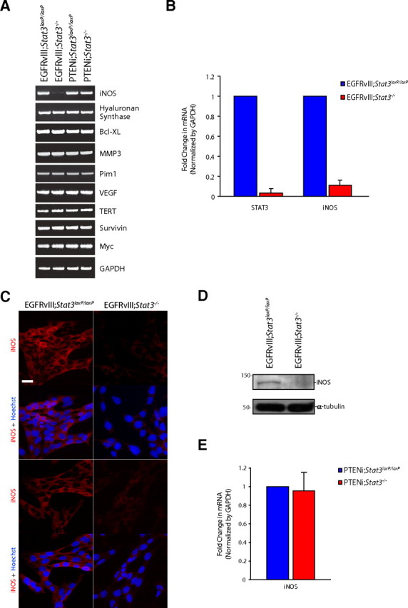
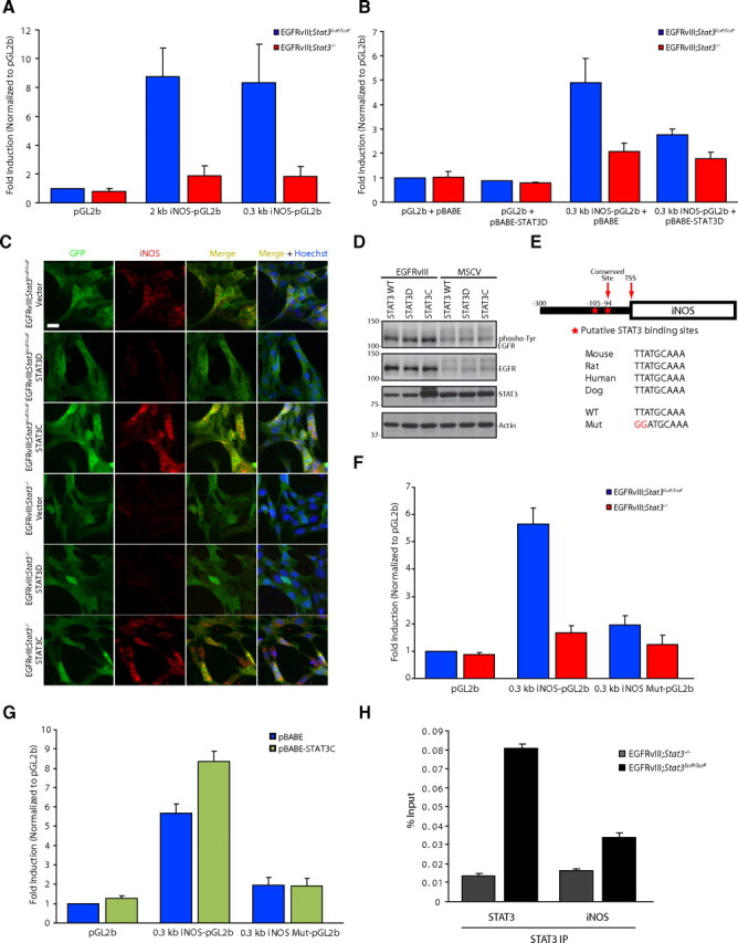
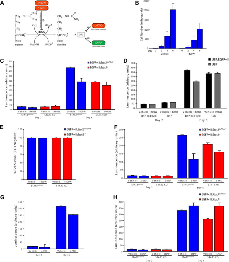
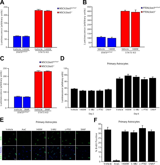
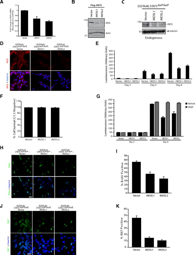
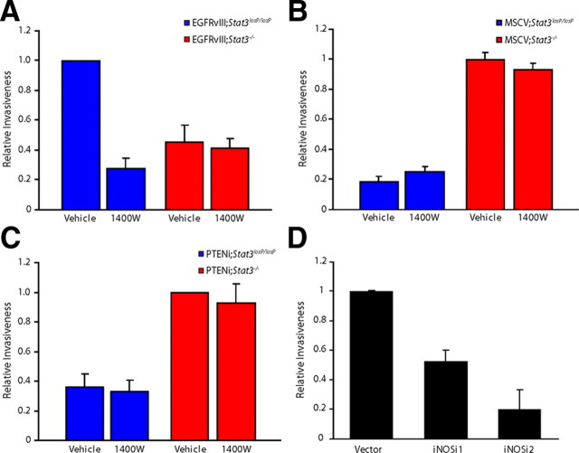
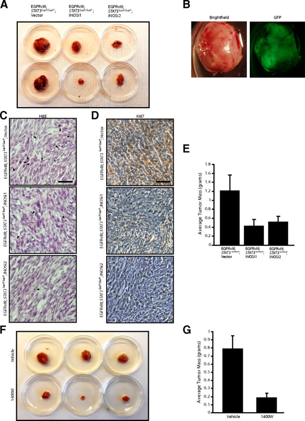
Malignant gliomas, including glioblastoma multiforme, constitute the most common and aggressive primary brain tumors in adults. The transcription factor signal transducer and activator of transcription 3 (STAT3) plays an essential role in glioblastoma pathogenesis downstream of the major oncogenic protein epidermal growth factor receptor variant III (EGFRvIII). However, the critical gene targets of STAT3 that mediate EGFRvIII-induced glial transformation have remained unknown. Here, we identify inducible nitric oxide synthase (iNOS) as a novel target gene of STAT3 in EGFRvIII-expressing mouse astrocytes. Endogenous STAT3 occupies the endogenous iNOS promoter and stimulates iNOS transcription in EGFRvIII-expressing astrocytes. STAT3 does not appear to control iNOS transcription in astrocytes deficient in the major glioblastoma tumor suppressor protein phosphatase and tensin homolog (PTEN), suggesting that STAT3 regulates iNOS transcription specifically in EGFRvIII-expressing astrocytes. Importantly, inhibition of iNOS by distinct approaches, including knockdown by RNA interference, reduces cell population growth and invasiveness of EGFRvIII-expressing astrocytes. In addition, upon iNOS knockdown or administration of a small-molecule inhibitor of iNOS, EGFRvIII-expressing astrocytes form smaller tumors in vivo. These findings suggest that inhibition of iNOS may have potential therapeutic value for EGFRvIII-activated brain tumors.


Figures

Figure 1.
Figure 1.
STAT3 regulates iNOS expression in EGFRvIII-expressing astrocytes. A, RNA was isolated from mouse EGFRvIII;Stat3loxP/loxP, EGFRvIII;Stat3−/−, PTENi;Stat3loxP/loxP, and PTENi;Stat3−/− astrocytes and subjected to RT-PCR with primers specific for the indicated genes. GAPDH served as control. iNOS mRNA levels were specifically reduced in EGFRvIII;Stat3−/− astrocytes compared with EGFRvIII;Stat3loxP/loxP astrocytes. B, RNA isolated from mouse EGFRvIII;Stat3loxP/loxP and EGFRvIII;Stat3−/− astrocytes was subjected to quantitative RT-PCR analyses using primers specific for STAT3 and iNOS. mRNA levels were normalized to GAPDH. STAT3 and iNOS mRNA levels were significantly reduced in EGFRvIII;Stat3−/− astrocytes compared with EGFRvIII;Stat3loxP/loxP astrocytes (ANOVA, p < 0.0005, n = 3). C, EGFRvIII;Stat3loxP/loxP and EGFRvIII;Stat3−/− astrocytes were subjected to immunocytochemistry using the rabbit iNOS antibody. Representative images are shown. The expression of iNOS was substantially reduced in EGFRvIII;Stat3−/− astrocytes compared with EGFRvIII;Stat3loxP/loxP astrocytes. Scale bar, 20 μm. D, Lysates of EGFRvIII;Stat3loxP/loxP and EGFRvIII;Stat3−/− astrocytes were immunoblotted with the iNOS or α-tubulin antibody. The levels of iNOS protein were substantially reduced in EGFRvIII;Stat3−/− astrocytes compared with EGFRvIII;Stat3loxP/loxP astrocytes. E, RNA isolated from mouse PTENi;Stat3loxP/loxP and PTENi;Stat3−/− astrocytes was subjected to quantitative RT-PCR analyses using primers specific for iNOS. mRNA levels were normalized to GAPDH. iNOS mRNA levels were not significantly different between PTENi;Stat3loxP/loxP astrocytes compared with PTENi;Stat3−/− astrocytes.
Figure 2.
Figure 2.
STAT3 directly regulates iNOS transcription in astrocytes. A, EGFRvIII;Stat3loxP/loxP and EGFRvIII;Stat3−/− astrocytes transfected with a luciferase reporter plasmid driven by a promoter containing the 2 or 0.3 kb region upstream of the iNOS transcriptional start site or the control pGL2-basic reporter plasmid together with a Renilla expression plasmid were subjected to dual luciferase assay 48 h after transfection. Expression of the luciferase reporter from both the 2 and 0.3 kb regions of the iNOS promoter was significantly increased in EGFRvIII;Stat3loxP/loxP astrocytes compared with EGFRvIII;Stat3−/− astrocytes (Kruskal–Wallis test, p < 0.01, n = 4). B, EGFRvIII;Stat3loxP/loxP and EGFRvIII;Stat3−/− astrocytes transfected with the 0.3 kb iNOS–luciferase reporter plasmid together with dominant-negative Stat3 (STAT3D) or the control vector and the Renilla expression plasmid were subjected to dual luciferase assay 48 h after transfection. Expression of the iNOS–luciferase reporter was significantly increased in EGFRvIII;Stat3loxP/loxP astrocytes compared with EGFRvIII;Stat3−/− astrocytes in the background of control vector. Expression of STAT3D significantly reduced the expression of the iNOS–luciferase reporter in EGFRvIII;Stat3loxP/loxP astrocytes (Kruskal–Wallis test, p < 0.005, n = 4). C, EGFRvIII;Stat3loxP/loxP and EGFRvIII;Stat3−/− astrocytes transfected with vector control, STAT3D, or STAT3C were subjected to immunocytochemistry using the GFP and iNOS antibodies. Endogenous iNOS expression was substantially reduced in EGFRvIII;Stat3loxP/loxP astrocytes upon expression of STAT3D and substantially increased in EGFRvIII;Stat3loxP/loxP and EGFRvIII;Stat3−/− astrocytes upon expression of STAT3C. Scale bar, 20 μm. D, Lysates of EGFRvIII-expressing and control MSCV-infected astrocytes transfected with wild-type Stat3 (STAT3 WT), STAT3D, or STAT3C were immunoblotted with the phospho-Tyr (4G10), EGFR, STAT3, or actin antibody. Expression of STAT3D or STAT3C had little or no effect on the tyrosine phosphorylation of EGFRvIII. E, Top, STAT3 binding site in iNOS 94 bp upstream of the transcriptional start site (TSS) is conserved. Bottom, Mutations introduced into the iNOS promoter to disrupt STAT3 binding. F, EGFRvIII;Stat3loxP/loxP and EGFRvIII;Stat3−/− astrocytes transfected with the 0.3 kb iNOS–luciferase or 0.3 kb iNOS–luciferase mutant reporter plasmid together with the Renilla expression plasmid were subjected to dual luciferase assay 48 h after transfection. Expression of the iNOS–luciferase reporter was significantly increased in EGFRvIII;Stat3loxP/loxP astrocytes compared with EGFRvIII;Stat3−/− astrocytes (Kruskal–Wallis test, p < 0.005, n = 5). Mutation of the STAT3 binding site significantly reduced iNOS-promoter-mediated expression in EGFRvIII;Stat3loxP/loxP astrocytes (Kruskal–Wallis test, p < 0.005, n = 5). G, EGFRvIII;Stat3loxP/loxP astrocytes transfected with the 0.3 kb iNOS–luciferase or 0.3 kb iNOS–luciferase mutant reporter plasmid together with STAT3C or the control vector and the Renilla expression plasmid were subjected to dual-luciferase assay 48 h after transfection. Expression of STAT3C significantly increased expression of the iNOS–luciferase reporter (Kruskal–Wallis test, p < 0.0005, n = 5) but had little or no effect on the expression of the iNOS–luciferase mutant reporter. H, EGFRvIII;Stat3loxP/loxP and EGFRvIII;Stat3−/− astrocytes were subjected to ChIP analyses using the STAT3 antibody. The STAT3 gene served as positive control. Endogenous STAT3 was significantly enriched at the endogenous iNOS promoter in EGFRvIII;Stat3loxP/loxP astrocytes compared with EGFRvIII;Stat3−/− astrocytes (ANOVA, p < 0.001, n = 3).
Figure 3.
Figure 3.
iNOS is required for the proliferation of EGFRvIII-expressing astrocytes. A, Schematic of NOS catalyzed by iNOS. Pharmacological agents used for analyses influence NO levels by distinct mechanisms. B, Population growth of EGFRvIII;Stat3loxP/loxP astrocytes exposed to vehicle or the iNOS inhibitor 1400W. Pharmacological inhibition of iNOS significantly reduced EGFRvIII-expressing astrocyte population growth (representative experiment of 3 independent experiments performed in triplicate; ANOVA, p < 0.0001, n = 3). C, Population growth of EGFRvIII;Stat3loxP/loxP and EGFRvIII;Stat3−/− astrocytes was assessed using an ATP-based assay of cell proliferation (Cell-Titer Glo). Pharmacological inhibition of iNOS with 1400W significantly reduced the population growth of EGFRvIII;Stat3loxP/loxP astrocytes (ANOVA, p < 0.0001, n = 5), with little or no effect on EGFRvIII;Stat3−/− astrocytes. D, Population growth of control and EGFRvIII-expressing U87 human glioblastoma cells was assessed as in C. Pharmacological inhibition of iNOS with 1400W significantly reduced the population growth of EGFRvIII-expressing U87 cells but had little or no effect on control U87 cells (representative experiment of 3 independent experiments performed in triplicate; ANOVA, p < 0.001, n = 3). E, EGFRvIII;Stat3loxP/loxP and EGFRvIII;Stat3−/− astrocytes were treated with the iNOS inhibitor 1400W and subjected to immunocytochemistry using the CC3 antibody. Exposure to 1400W had little or no effect on the percentage of cells that were CC3 negative in both EGFRvIII;Stat3loxP/loxP and EGFRvIII;Stat3−/− astrocytes. F, Population growth of EGFRvIII;Stat3loxP/loxP and EGFRvIII;Stat3−/− astrocytes was assessed as in C. Pharmacological inhibition of iNOS with S-MIU significantly reduced the population growth of EGFRvIII;Stat3loxP/loxP astrocytes (ANOVA, p < 0.0001, n = 3), with a more modest effect on EGFRvIII;Stat3−/− astrocytes. G, Population growth of EGFRvIII;Stat3loxP/loxP and EGFRvIII;Stat3−/− astrocytes was assessed as in C. The NO scavenger c-PTIO significantly reduced the population growth of EGFRvIII;Stat3loxP/loxP astrocytes (ANOVA, p < 0.0001, n = 3). H, Population growth of EGFRvIII;Stat3loxP/loxP and EGFRvIII;Stat3−/− astrocytes was assessed as in C. The NO donor SNAP modestly increased the population growth of EGFRvIII;Stat3loxP/loxP astrocytes and significantly increased the population growth of EGFRvIII;Stat3−/− astrocytes (ANOVA, p < 0.0001, n = 3). KO, Knock-out.
Figure 4.
Figure 4.
iNOS is not required for the proliferation of control and PTEN-deficient astrocytes. A, Population growth of MSCV;Stat3loxP/loxP and MSCV;Stat3−/− astrocytes was assessed as in Figure 3C. Pharmacological inhibition of iNOS with 1400W had little or no effect on the population growth of MSCV;Stat3loxP/loxP and MSCV;Stat3−/− astrocytes compared with vehicle treatment (n = 3). B, Population growth of PTENi;Stat3loxP/loxP and PTENi;Stat3−/− astrocytes was assessed as in Figure 3C. Pharmacological inhibition of iNOS with 1400W had little or no effect on the population growth of PTENi;Stat3loxP/loxP and PTENi;Stat3−/− astrocytes compared with vehicle treatment (n = 3). C, Population growth of MSCV;Stat3loxP/loxP and MSCV;Stat3−/− astrocytes was assessed as in Figure 3C. The NO donor SNAP had little or no effect on the population growth of MSCV;Stat3loxP/loxP and MSCV;Stat3−/− astrocytes compared with vehicle treatment (n = 3). D, Population growth of primary mouse astrocytes was assessed as in Figure 3C. Pharmacological inhibitors and activators of the iNOS pathway had little or no effect on the population growth of primary mouse astrocytes compared with vehicle treatment (n = 3). E, Primary mouse astrocytes treated with inhibitors and activators of the iNOS pathway were subjected to immunocytochemistry using the BrdU antibody. Inhibition or activation of the iNOS pathway had little or no effect on BrdU incorporation of primary mouse astrocytes compared with vehicle treatment. In control experiments, the nucleoside analog AraC substantially reduced BrdU incorporation in primary astrocytes compared with vehicle treatment. Scale bar, 20 μm. F, Primary mouse astrocytes analyzed as in E were quantified for the percentage of BrdU-positive cells. Inhibition or activation of the iNOS pathway had little or no effect on BrdU incorporation in primary astrocytes. In control experiments, the nucleoside analog AraC significantly reduced BrdU incorporation compared with vehicle treatment (ANOVA, p < 0.0001, n = 3). KO, Knock-out.
Figure 5.
Figure 5.
iNOS knockdown reduces the proliferation of EGFRvIII-expressing astrocytes. A, RNA isolated from mouse EGFRvIII;Stat3loxP/loxP astrocytes infected with iNOS RNAi virus or control virus was subjected to quantitative RT-PCR analyses using primers specific for iNOS. mRNA levels were normalized to GAPDH. iNOS mRNA levels were significantly reduced in EGFRvIII;Stat3loxP/loxP iNOS knockdown astrocytes compared with vector-transfected astrocytes (ANOVA, p < 0.005, n = 4). B, Lysates of HEK293T cells transfected with an expression plasmid encoding Flag–iNOS together with the iNOS RNAi or control RNAi plasmid were immunoblotted with the Flag or actin antibody. Knockdown of iNOS reduced the levels of iNOS protein in HEK293T cells. C, Lysates of control and iNOS knockdown EGFRvIII;Stat3loxP/loxP astrocytes were immunoblotted with the iNOS or α-tubulin antibody. Knockdown of iNOS substantially reduced the levels of endogenous iNOS protein in astrocytes. D, Control and iNOS knockdown EGFRvIII;Stat3loxP/loxP astrocytes were subjected to immunocytochemistry using the iNOS antibody. Representative images are shown. The expression of endogenous iNOS protein was substantially reduced in iNOS knockdown astrocytes compared with control astrocytes. Scale bar, 20 μm. E, Population growth of control and iNOS knockdown EGFRvIII;Stat3loxP/loxP astrocytes was assessed as in Figure 3C. iNOS knockdown significantly reduced the population growth of EGFRvIII;Stat3loxP/loxP astrocytes (ANOVA, p < 0.0001, n = 3). F, Control and iNOS knockdown EGFRvIII;Stat3loxP/loxP astrocytes were subjected to immunocytochemistry using the CC3 antibody. Knockdown of iNOS had little or no effect on the percentage of astrocytes that were CC3 negative (n = 3). G, Population growth of control and iNOS knockdown EGFRvIII;Stat3loxP/loxP astrocytes was assessed as in Figure 3C. The NO donor SNAP significantly increased the population growth of iNOS knockdown EGFRvIII;Stat3−/− astrocytes (representative experiment of 3 independent experiments performed in triplicate; ANOVA, p < 0.001, n = 3). H, Control and iNOS knockdown EGFRvIII;Stat3loxP/loxP astrocytes were subjected to immunocytochemistry using the BrdU antibody. Incorporation of BrdU in astrocytes was substantially reduced upon iNOS knockdown. Scale bar, 20 μm. I, Control and iNOS knockdown EGFRvIII;Stat3loxP/loxP astrocytes analyzed as in H were quantified for the percentage of BrdU-positive cells. Incorporation of BrdU was substantially reduced in iNOS knockdown astrocytes compared with control astrocytes (ANOVA, p < 0.0001, n = 3). J, Control and iNOS knockdown EGFRvIII;Stat3loxP/loxP astrocytes were subjected to immunocytochemistry using the Ki67 antibody. Immunoreactivity of Ki67 was substantially reduced in iNOS knockdown astrocytes. Scale bar, 20 μm. K, Control and iNOS knockdown EGFRvIII;Stat3loxP/loxP astrocytes analyzed as in J were quantified for the percentage of Ki67-positive cells. The percentage of Ki67-positive cells was significantly reduced in iNOS knockdown astrocytes compared with control astrocytes (ANOVA, p < 0.0001, n = 3).
Figure 6.
Figure 6.
iNOS knockdown impairs the invasiveness of EGFRvIII-expressing astrocytes. A, EGFRvIII;Stat3loxP/loxP and EGFRvIII;Stat3−/− astrocytes were seeded on top of an 8 μm pore size insert coated with matrigel in the presence of 1400W or control vehicle and allowed to invade through the matrigel matrix for 22 h. 1400W treatment significantly reduced the invasiveness of EGFRvIII;Stat3loxP/loxP astrocytes (ANOVA, p < 0.005, n = 3) but had little or no effect on EGFRvIII;Stat3−/− astrocytes. The effect of 1400W treatment on invasiveness was not secondary to a change in cell proliferation, because the invasive potential of these cells was measured at a time (22 h after plating) before a significant decrease in cell number upon iNOS inhibition. B, Invasiveness of MSCV;Stat3loxP/loxP and MSCV;Stat3−/− astrocytes exposed to 1400W was assessed as in A. Exposure to 1400W had little or no effect on the invasiveness of MSCV;Stat3loxP/loxP and MSCV;Stat3−/− astrocytes compared with vehicle treatment (n = 3). C, Invasiveness of PTENi;Stat3loxP/loxP and PTENi;Stat3−/− astrocytes exposed to 1400W was assessed as in A. Exposure to 1400W had little or no effect on the invasiveness of PTENi;Stat3loxP/loxP and PTENi;Stat3−/− astrocytes compared with vehicle treatment (n = 3). D, Invasiveness of control and iNOS knockdown EGFRvIII;Stat3loxP/loxP astrocytes was assessed as in A. Knockdown of iNOS significantly reduced EGFRvIII;Stat3loxP/loxP astrocyte cell invasiveness (ANOVA, p < 0.005, n = 4). The effect of iNOS knockdown on invasiveness was not secondary to a change in cell proliferation, because the invasive potential of these cells was measured at a time (22 h after plating) before a significant decrease in cell number upon iNOS knockdown.
Figure 7.
Figure 7.
iNOS is required for the ability of EGFRvIII-expressing astrocytes to form tumors in vivo. A, Control and iNOS knockdown EGFRvIII;Stat3loxP/loxP astrocytes were injected subcutaneously into SCID mice. Four weeks after injection, tumors were removed, measured, and stained. iNOS knockdown reduced tumor size. B, Representative excised subcutaneous tumor revealed that masses removed were GFP positive and derived from injected cells. C, Tumors derived from control and iNOS knockdown EGFRvIII;Stat3loxP/loxP astrocytes were analyzed by hematoxylin and eosin (H&E) staining. Control EGFRvIII;Stat3loxP/loxP astrocytes showed histological features of neoplastic transformation, including nuclear atypia, hypercellularity, and frequent mitotic figures (arrowheads), although these features were less prominent in iNOS knockdown tumors. Scale bar, 200 μm. D, Tumors derived from control and iNOS knockdown EGFRvIII;Stat3loxP/loxP astrocytes were analyzed by immunohistochemical analyses using the Ki67 antibody. Tumors derived from control EGFRvIII;Stat3loxP/loxP astrocytes had substantially greater Ki67 immunoreactivity compared with tumors derived from iNOS knockdown EGFRvIII;Stat3loxP/loxP astrocytes. Scale bar, 200 μm. E, Average tumor mass of tumors from A was measured. iNOS knockdown significantly reduced the mass of EGFRvIII;Stat3loxP/loxP tumors (ANOVA, p < 0.05, n = 33 animals). F, EGFRvIII;Stat3loxP/loxP astrocytes were injected subcutaneously into SCID mice, and the iNOS inhibitor 1400W or vehicle control was locally administered. Four weeks after initial injections, tumors were removed, measured, and stained. Treatment with the iNOS inhibitor 1400W substantially reduced tumor size. G, Average tumor mass of tumors from F was measured. Treatment with the iNOS inhibitor 1400W substantially reduced the mass of EGFRvIII;Stat3loxP/loxP tumors (t test, p < 0.005, n = 20 animals). Two animals in the 1400W-treated cohort did not have detectable tumors at the endpoint.

Similar articles

Cited by

References

    1. Akaike T, Yoshida M, Miyamoto Y, Sato K, Kohno M, Sasamoto K, Miyazaki K, Ueda S, Maeda H. Antagonistic action of imidazolineoxyl N-oxides against endothelium-derived relaxing factor/.NO through a radical reaction. Biochemistry. 1993;32:827–832. - PubMed
    1. Aoki Y, Feldman GM, Tosato G. Inhibition of STAT3 signaling induces apoptosis and decreases survivin expression in primary effusion lymphoma. Blood. 2003;101:1535–1542. - PubMed
    1. Bachoo RM, Maher EA, Ligon KL, Sharpless NE, Chan SS, You MJ, Tang Y, DeFrances J, Stover E, Weissleder R, Rowitch DH, Louis DN, DePinho RA. Epidermal growth factor receptor and Ink4a/Arf: convergent mechanisms governing terminal differentiation and transformation along the neural stem cell to astrocyte axis. Cancer Cell. 2002;1:269–277. - PubMed
    1. Bajenaru ML, Hernandez MR, Perry A, Zhu Y, Parada LF, Garbow JR, Gutmann DH. Optic nerve glioma in mice requires astrocyte Nf1 gene inactivation and Nf1 brain heterozygosity. Cancer Res. 2003;63:8573–8577. - PubMed
    1. Bonni A, Sun Y, Nadal-Vicens M, Bhatt A, Frank DA, Rozovsky I, Stahl N, Yancopoulos GD, Greenberg ME. Regulation of gliogenesis in the central nervous system by the JAK-STAT signaling pathway. Science. 1997;278:477–483. - PubMed

Publication types

MeSH terms