. 2012 Jun 6;32(23):7895-906.
doi: 10.1523/JNEUROSCI.0445-12.2012.

Irregular Breathing in Mice following Genetic Ablation of V2a Neurons

Affiliations

Irregular Breathing in Mice following Genetic Ablation of V2a Neurons

Steven A Crone et al. J Neurosci. .

Abstract

Neural networks called central pattern generators (CPGs) generate repetitive motor behaviors such as locomotion and breathing. Glutamatergic neurons are required for the generation and inhibitory neurons for the patterning of the motor activity associated with repetitive motor behaviors. In the mouse, glutamatergic V2a neurons coordinate the activity of left and right leg CPGs in the spinal cord enabling mice to generate an alternating gait. Here, we investigate the role of V2a neurons in the neural control of breathing, an essential repetitive motor behavior. We find that, following the ablation of V2a neurons, newborn mice breathe at a lower frequency. Recordings of respiratory activity in brainstem-spinal cord and respiratory slice preparations demonstrate that mice lacking V2a neurons are deficient in central respiratory rhythm generation. The absence of V2a neurons in the respiratory slice preparation can be compensated for by bath application of neurochemicals known to accelerate the breathing rhythm. In this slice preparation, V2a neurons exhibit a tonic firing pattern. The existence of direct connections between V2a neurons in the medial reticular formation and neurons of the pre-Bötzinger complex indicates that V2a neurons play a direct role in the function of the respiratory CPG in newborn mice. Thus, neurons of the embryonic V2a lineage appear to have been recruited to neural networks that control breathing and locomotion, two prominent CPG-driven, repetitive motor behaviors.


Figures
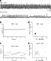
Figure 1.
Figure 1.
Ablation of V2a neurons in the medulla of Chx10::DTA mice results in slow, irregular breathing. a, Whole-body plethysmographs from P0 wild-type (top) and Chx10::DTA (bottom) mice born to C57BL/6 dams show inspiration as upward deflections in the graph. b, c, Instantaneous respiratory frequency (breaths/minute) is plotted for 50 consecutive breaths in a P0 wild-type mouse (b) and a Chx10::DTA mouse (c) born to C57BL/6 dams to illustrate the breath to breath consistency of respiratory frequency. d, e, Poincaré maps of the respiratory period (Tn) in seconds versus the subsequent period (Tn+1) of a P0 wild type (d) and a Chx10::DTA (e) mouse as measured by plethysmography.
Figure 2.
Figure 2.
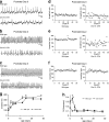
Postnatal changes in breathing regularity by Chx10::DTA mice. a–c, Whole-body plethysmography was performed at P0 (a), P2 (b), and P4 (c) on the same Chx10::DTA mouse (bottom) and wild-type littermate (top) born to an ICR dam. d–f, Instantaneous respiratory frequency (breaths/minute) is plotted for 50 consecutive breaths at ages P0 (d), P2 (e), and P4 (f) from the same wild-type mouse (left) and Chx10::DTA mouse (right). g, Respiratory frequency as a function of age in P0–P28 wild-type and Chx10::DTA mice. h, The CV of the respiratory frequency as a function of age in P0–P28 wild-type and Chx10::DTA mice. g, h, Error bars indicate SEM. The asterisk (*) denotes Chx10::DTA versus wild type. p < 0.05 by Student's t test (g) or Mann–Whitney rank sum test (h). The number of wild-type and Chx10::DTA animals, respectively, analyzed for each age is P0 (4, 6), P2 (4, 5), P4 (4, 5), P6 (4, 5), P12 (4, 3), P15 (3, 3), P21 (5, 5), and P28 (6, 6).
Figure 3.
Figure 3.
Respiratory rhythm generation is slow and irregular in P0 Chx10::DTA mice. a–d, Activity of the C4 ventral root was recorded in brainstem–spinal cord preparations from wild-type (a) and Chx10::DTA (b) mice. Integrated (top trace) and raw (bottom trace) C4 root recordings are shown. The average frequency in hertz (c) of C4-root burst activity is significantly decreased, whereas the variability (coefficient of variation) of the interburst interval (d) is increased in Chx10::DTA (n = 7) compared with control mice (n = 6). e, f, Coronal sections of age P0 medulla from wild-type (e) and Chx10::DTA (f) mice immunostained for NK1R (red) and ChAT (green). NK1R marks neurons of the pre-BötC and nucleus ambiguus, whereas ChAT marks only the nucleus ambiguus. g, h, At E15.5, the medulla of a Chx10::DTA (h) embryo shows a lack of Chx10 neurons (green), but no changes in NK1R (red) staining compared with a wild-type (g) embryo. i, j, Rectified (top) and raw (bottom) extracellular recordings of rhythmic burst activity in the VRG of medullary slice preparations from P0 wild-type (i) and Chx10::DTA (j) mice. k, The average frequency in hertz of VRG burst activity in medullary slices from wild-type (n = 10) and Chx10::DTA mice (n = 10). Decreased burst frequency in Chx10::DTA mice compared with wild type is accompanied by higher variability in the interburst interval [measured as the coefficient of variation (l)]. Error bars indicate SEM. m, n, Poincaré maps of the period between bursts (Tn) in seconds versus the subsequent period (Tn+1) of VRG burst activity in medullary slices from a P0 wild-type (m) and a Chx10::DTA (n) mouse. Scale bars: e, f, 50 μm; g, h, 100 μm.
Figure 4.
Figure 4.
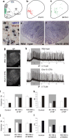
Glutamatergic V2a neurons expressing Chx10 are found in the mRF of the medulla. a, Schematic outline of a coronal hemisection through the medulla of an adult mouse brain. Structures containing respiratory related neurons are shown: hypo-glossal (XII), vagus (X), and nucleus ambiguus (NA) motor nuclei, nucleus of the solitary tract (NTS), raphe, medial reticular formation (mRF), and the pre-BötC. Also shown is the inferior olivary nucleus (IO). b, c, Drawings of coronal sections from age P0 mouse medulla showing the location of Chx10 (b) and GATA3 (c) interneurons as determined by immunohistochemistry. Chx10 neurons are located predominantly within the presumed mRF region. d, Dual in situ hybridization and immunohistochemistry in the medullary mRF demonstrate that Chx10 neurons (brown nuclei) express vglut2 mRNA (blue cytoplasmic staining). e, f, In situ hybridization for vglut2 mRNA at age P0 shows fewer glutamatergic neurons in Chx10::DTA (f) compared with wild-type (e) mice in the mRF (outlined by dashed line), but not in dorsolateral medulla. g, The distribution of SP immunoreactivity is similar in the medulla of P0 wild-type (top) and Chx10::DTA mice (bottom). h, Rectified traces showing that bath-applied SP (0.75 μm) increases the burst frequency of the VRG in medullary slices of P0 wild-type (top) and Chx10::DTA mice (bottom). The gray box marks the period between 60 and 180 s after SP application. i, k, The effects of exogenous SP on frequency (i) and coefficient of variation of the interburst interval (k) in medullary slices from wild-type (white bars; n = 5) and Chx10::DTA (black bars; n = 5) mice before SP application (control), between 60 and 180 s of SP (gray box), and between 180 and 500 s of SP. j, l, The effects of exogenous NMDA on frequency (j) and coefficient of variation of the interburst interval (l) in medullary slices from wild-type (white bars; n = 4) and Chx10::DTA (black bars; n = 4) mice before NMDA application (control), between 60 and 180 s of NMDA (gray box), and between 180 and 500 s of NMDA. Error bars indicate SEM. Scale bars: d, 20 μm; e–g, 100 μm.
Figure 5.
Figure 5.
Firing properties of fluorescently labeled V2a neurons. a, Coronal section from a P0 Chx10::CFP mouse at the level of the pre-BötC. The soma of CFP-labeled V2a neurons are restricted to the mRF. b–d, Whole-cell current-clamp recordings of CFP expressing V2a neurons in medullary slices made from neonatal Chx10::CFP mice. b, V2a neurons exhibit a strong inward rectification (Ih) when hyperpolarized below −150 mV. The inset shows current step protocol used to inject 50 pA steps from −150 to +100 pA. c, Relationship between current injection and mean firing rate in V2a neurons (n = 4 cells). Depolarization block was observed above a maximum firing frequency of 50 Hz. Error bars indicate SEM. d, Spontaneous tonic firing activity of a V2a neuron at a resting membrane potential of −62 mV. Raising the extracellular [K]o from 3 to 5 mm causes an increase in firing frequency but the firing pattern remains tonic. Scale bar, 100 μm.
Figure 6.
Figure 6.
V2a neurons project to the pre-BötC. a–c, Coronal sections of the medulla from a P0 Chx10::CFP mouse stained with ChAT (green) and NK1R (red). The box in a is shown at higher magnification in b (NK1R only) and c (ChAT and NK1R). The NA forms a cluster of large neurons that are double labeled by ChAT and NK1R (c). The arrows mark some of the NK1R+ neurons located below the NA in the pre-BötC. d–f, An adjacent section is immunostained for CFP (green) and NK1R (red), with the relative location of the NA in a and c shown with a circle in d and f. The box in d is shown at higher magnification in e (NK1R only) and f (CFP and NK1R). The arrows mark some of the NK1R+ neurons below the NA in the pre-BötC. CFP+ fibers from V2a neurons project to the pre-BötC region below the NA. Scale bar, 50 μm.
Figure 7.
Figure 7.
V2a neurons make vGLUT2-enriched synapses in the pre-BötC. a, b, Retrograde labeling of V2a neurons projecting to the VRG. The VRG was identified using electrophysiology based on rhythmic burst activity in medullary slices, and fluorescein-dextran (green) was injected to retrogradely label neurons projecting to this region. The green pipette drawing marks the site of injection in a cryostat section from the physiological slice preparation (a). V2a neurons were identified using antibodies to Chx10 (red). The area of the mRF marked by a rectangle is shown at higher magnification in b. Many neurons in the mRF can be retrogradely labeled (green) from the pre-BötC, including Chx10 neurons (yellow). Some Chx10 neurons are not retrogradely labeled (red). c–h, Confocal images of the pre-BötC from P0 Chx10::CFP mice showing excitatory projections from V2a neurons onto the processes (c–g) and cell bodies (h–l) of NK1R+ neurons. NK1R immunoreactivity (red) marks the cell bodies and processes of specific neurons in the pre-BötC. vGLUT2 (blue) marks excitatory synaptic terminals and CFP (green) marks the processes of V2a neurons. The boxed areas (c, h) are shown at higher magnification in d–g and i–l. Excitatory terminals from V2a neurons are indicated in white as overlap between green and blue (g, l). The arrows in higher magnification images indicate V2a excitatory terminals that are in contact with NK1R processes (d–g) or cell body (i–l). Scale bars: a, b, 50 μm; c–l, 5 μm.

Similar articles

Cited by

References

    1. Alheid GF, McCrimmon DR. The chemical neuroanatomy of breathing. Respir Physiol Neurobiol. 2008;164:3–11. - PMC - PubMed
    1. Al-Mosawie A, Wilson JM, Brownstone RM. Heterogeneity of V2-derived interneurons in the adult mouse spinal cord. Eur J Neurosci. 2007;26:3003–3015. - PubMed
    1. Amiel J, Dubreuil V, Ramanantsoa N, Fortin G, Gallego J, Brunet JF, Goridis C. PHOX2B in respiratory control: lessons from congenital central hypoventilation syndrome and its mouse models. Respir Physiol Neurobiol. 2009;168:125–132. - PubMed
    1. Borday C, Chatonnet F, Thoby-Brisson M, Champagnat J, Fortin G. Neural tube patterning by Krox20 and emergence of a respiratory control. Respir Physiol Neurobiol. 2005;149:63–72. - PubMed
    1. Bouvier J, Thoby-Brisson M, Renier N, Dubreuil V, Ericson J, Champagnat J, Pierani A, Chédotal A, Fortin G. Hindbrain interneurons and axon guidance signaling critical for breathing. Nat Neurosci. 2010;13:1066–1074. - PubMed

Publication types

MeSH terms