. 2012 Jun 6;32(23):8074-83.
doi: 10.1523/JNEUROSCI.0129-12.2012.

Prolactin regulates tuberoinfundibular dopamine neuron discharge pattern: novel feedback control mechanisms in the lactotrophic axis

Affiliations

Prolactin regulates tuberoinfundibular dopamine neuron discharge pattern: novel feedback control mechanisms in the lactotrophic axis

David J Lyons et al. J Neurosci. .

Abstract

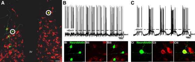
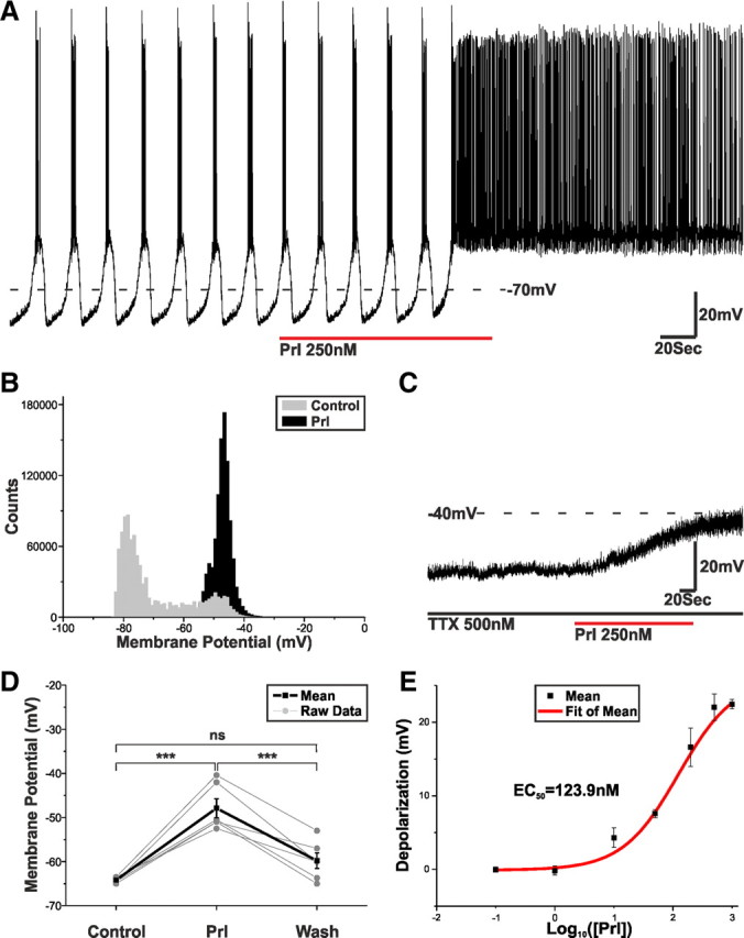
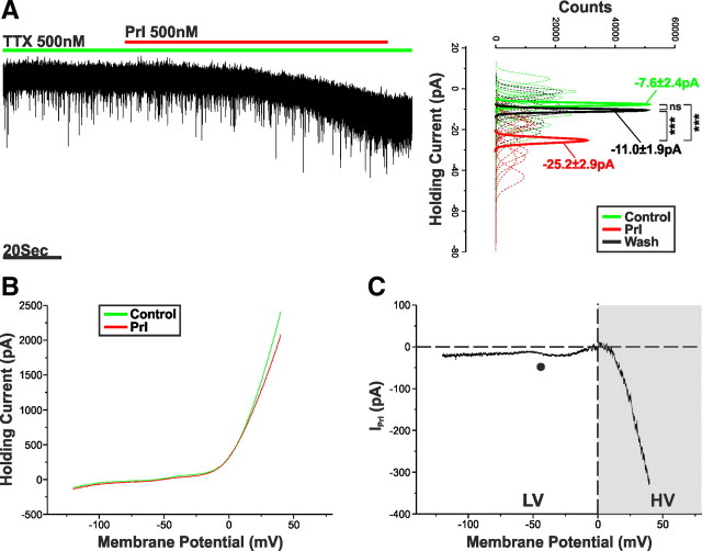
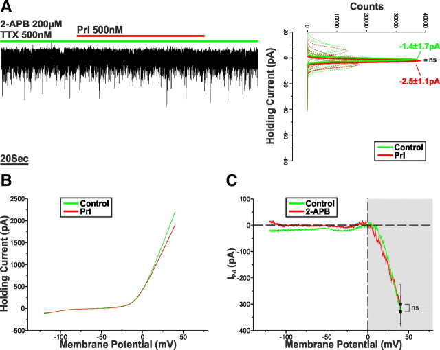
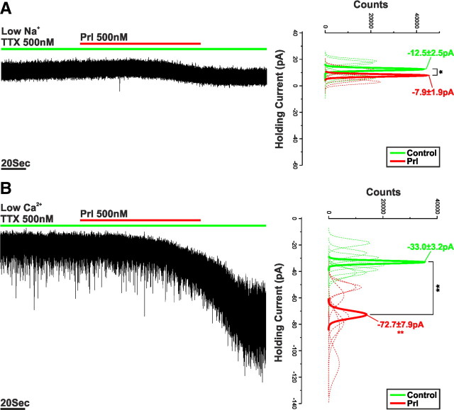
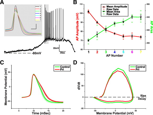
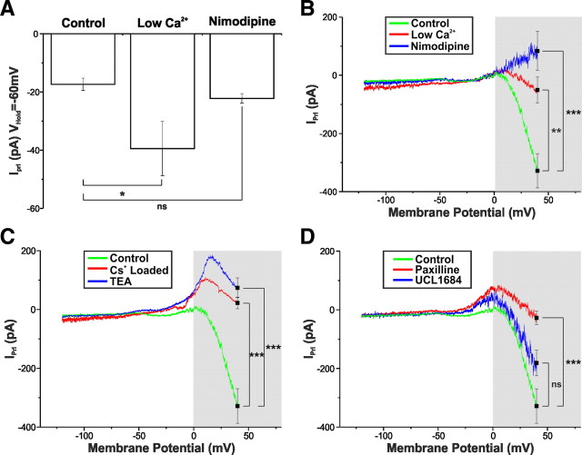
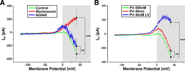
Balance in the body's hormonal axes depends on feedback onto neuroendocrine hypothalamic neurons. This phenomenon involves transcriptional and biosynthetic effects, yet less is known about the potential rapid modulation of electrical properties. Here, we investigated this issue in the lactotrophic axis, in which the pituitary hormone prolactin is tonically inhibited by tuberoinfundibular dopamine (TIDA) neurons located in the hypothalamic arcuate nucleus. Whole-cell recordings were performed on slices of the rat hypothalamus. In the presence of prolactin, spontaneously oscillating TIDA cells depolarized, switched from phasic to tonic discharge, and exhibited broadened action potentials. The underlying prolactin-induced current is composed of separate low- and high-voltage components that include the activation of a transient receptor potential-like current and the inhibition of a Ca(2+)-dependent BK-type K(+) current, respectively, as revealed by ion substitution experiments and pharmacological manipulation. The two components of the prolactin-induced current appear to be mediated through distinct signaling pathways as the high-voltage component is abolished by the phosphoinositide 3-kinase blocker wortmannin, whereas the low-voltage component is not. This first description of the central electrophysiological actions of prolactin suggests a novel feedback mechanism. By simultaneously enhancing the discharge and spike duration of TIDA cells, increased serum prolactin can promote dopamine release to limit its own secretion with implications for the control of lactation, sexual libido, fertility, and body weight.


Figures

Figure 1.
Figure 1.
TIDA neurons display rhythmic oscillations. A, Merged confocal stack of a 250-μm-thick hypothalamic Arc slice processed with avidin–fluorescein to detect recorded Neurobiotin-filled neurons (green) and stained with immunofluorescence for TH (red) to identify TIDA neurons. Of the four recorded neurons in the left side of the third ventricle (3V), none displayed oscillatory behavior or stained positive for TH immunofluorescence. Scale bar, 50 μm. B, Current-clamp recording of the circled Neurobiotin-filled neuron from the left side of the 3V; high-magnification micrographs of this neuron shown in Bi–Biii. Note the lack of oscillatory discharge and absence of TH immunoreactivity. C, Current-clamp recording of the circled Neurobiotin-filled neuron from the right side of the 3V; high-magnification micrographs of this neuron shown in Ci–Ciii. Scale bar, 10 μm. Note robust regular membrane potential oscillation and colocalization of Neurobiotin and TH immunoreactivity.
Figure 2.
Figure 2.
Prl switches TIDA discharge from phasic to tonic. A, Current-clamp recording of an oscillating TIDA neuron. Application of Prl switches TIDA discharge from phasic to tonic. B, Frequency distribution plot demonstrating the Prl-induced shift in membrane potential from a biphasic to a monophasic distribution. C, Current-clamp recording of an oscillating TIDA neuron. The Prl-induced depolarization endures in the presence of TTX. D, Prl (250 nm) causes a reversible and significant change in resting membrane potential (recorded in TTX and from an initial membrane potential of −65 mV, a value achieved though the injection of negative direct holding current). ***p < 0.001. E, Dose–response curve highlighting the relationship between Prl concentration and the resultant membrane depolarization. Cells included were exposed to single concentrations of Prl in the presence of TTX and from a resting membrane potential of −65 mV.
Figure 3.
Figure 3.
Prl activates an inward current. A, Voltage-clamp recording of an oscillating TIDA neuron in the presence of TTX. Application of Prl results in an inward current. To the right, sharing its y-axis with the raw trace, are Gaussian fits of averaged (solid lines) holding current frequency distributions in control (green), Prl (red), and wash (black). Raw data used to produce averages shown as dashed lines. ***p < 0.001. B, Averaged voltage-clamp ramps (n = 14) acquired in control (green) and at the peak of the Prl response (red). C, Prl-induced current obtained by the digital subtraction of the traces displayed in B. Note the negative slope conductance at −50 mV (●), lack of reversal, and increased inward current in the HV range (gray box).
Figure 4.
Figure 4.
The LV component of IPrl is abolished by 2-APB. A, Voltage-clamp recording of an oscillator neuron in the presence of TTX and 2-APB (200 μm). Application of Prl failed to induce an inward current. To the right, sharing its y-axis with the raw trace, are Gaussian fits of averaged (solid lines) holding current frequency distributions in control (green) and Prl (red). Raw data used to produce averages shown as dashed lines. B, Averaged voltage-clamp ramps recorded in the presence of 2-APB (200 μm; n = 9) acquired in control (green) and at the peak of the Prl response (red). C, Prl-induced current obtained by the digital subtraction of the traces displayed in B. Note the marked reduction in inward current throughout the LV but not HV range.
Figure 5.
Figure 5.
The LV component of IPrl is mediated by a Na+-dominated MCC. A, Voltage-clamp recording of an oscillating TIDA neuron in the presence of TTX and zero-Na+ recording solution. Application of Prl induced an inward current that was significantly reduced when compared with control. To the right, sharing its y-axis with the raw trace, are Gaussian fits of averaged (solid lines) holding current frequency distributions in control (green) and Prl (red). Raw data used to produce averages shown as dashed lines (n = 5). B, Voltage-clamp recording of an oscillating TIDA neuron in the presence of TTX and low-Ca2+/high-Mg2+ recording solution. Application of Prl induced an inward current that was significantly enhanced when compared with control. To the right, sharing its y-axis with the raw trace, are Gaussian fits of averaged (solid lines) holding current frequency distributions in control (green) and Prl (red). Raw data used to produce averages shown as dashed lines (n = 7). *p < 0.05, **p < 0.01.
Figure 6.
Figure 6.
Prl broadens TIDA APs. A, Current-clamp recording of a TIDA neuron up state. Gray inset, An expanded superimposition of the first seven peak-aligned APs demonstrating variation in waveform properties. B, Mean ± SEM of the AP amplitude (red) and AP area (green) of the first seven APs of five consecutive up states from five different TIDA neurons. Amplitude and area achieved statistical significance compared with AP1 from AP2 onward. C, Average AP waveforms from a TIDA neuron in control (green) and Prl (red; 500 nm; n = 10). Raw data used to produce averages shown as dashed lines. To control for the variation in AP properties, only groups with statistically similar amplitudes and thresholds were used for analysis. Note the Prl-induced broadening of the AP. D, Phase plots of the average AP waveforms depicted in C. Note the Prl-induced changes in rise and decay.
Figure 7.
Figure 7.
The HV component of IPrl is underpinned by the inhibition of a Ca2+-sensitive BK-type K+ current. A, Summary of the effects of low-Ca2+/high-Mg2+ solution (n = 7) and nimodipine (10 μm; n = 5) on IPrl at VHold = −60 mV to control (n = 11). Low-Ca2+/high-Mg2+ solution significantly augments the Prl-induced inward current. Values expressed as mean ± SEM. *p < 0.05. B, I–V relationship of IPrl in control (green, n = 14), low-Ca2+/high-Mg2+ solution (red; n = 7), and nimodipine (10 μm; blue; n = 5). Both nimodipine and low-Ca2+/high-Mg2+ solution significantly modify IPrl at VHold = +40 mV. Values expressed as mean ± SEM. **p < 0.01, ***p < 0.005. C, I–V relationship of IPrl in control (green; n = 14), “Cs+-loaded” solution (red; n = 6), and TEA (10 mm; blue; n = 7). Both Cs+-loaded solution and TEA significantly modified IPrl at VHold = +40 mV. Values expressed as mean ± SEM. ***p < 0.005. D, I–V relationship of IPrl in control (green; n = 14), paxilline (2 μm; red; n = 8), and UCL1684 (1 μm; blue; n = 6). Paxilline, but not UCL1684, significantly modified IPrl at VHold = +40 mV. Values expressed as mean ± SEM. ***p < 0.005.
Figure 8.
Figure 8.
The LV and HV components of IPrl are activated by distinct signal transduction pathways. A, I–V relationship of IPrl in control (green; n = 14), wortmannin (200 nm; red; n = 8), and AG490 (10 μm; blue; n = 3). Wortmannin, but not AG490, significantly modifies IPrl at VHold = +40 mV. Values expressed as mean ± SEM. ***p < 0.005. B, I–V relationship of IPrl at a concentration of 500 nm Prl (green; n = 14), 40 nm Prl in which both the LV and HV components were present (red; n = 13 of 19), and 40 nm Prl in which only the LV component was present (blue; n = 6 of 19). Values expressed as mean ± SEM. ***p < 0.005.

References

    1. al-Sakkaf KA, Dobson PR, Brown BL. Activation of phosphatidylinositol 3-kinase by prolactin in Nb2 cells. Biochem Biophys Res Commun. 1996;221:779–784. - PubMed
    1. Amenomori Y, Chen CL, Meites J. Serum prolactin levels in rats during different reproductive states. Endocrinology. 1970;86:506–510. - PubMed
    1. Andrews ZB, Kokay IC, Grattan DR. Dissociation of prolactin secretion from tuberoinfundibular dopamine activity in late pregnant rats. Endocrinology. 2001;142:2719–2724. - PubMed
    1. Arbogast LA, Voogt JL. Hyperprolactinemia increases and hypoprolactinemia decreases tyrosine hydroxylase messenger ribonucleic acid levels in the arcuate nuclei, but not the substantia nigra or zona incerta. Endocrinology. 1991;128:997–1005. - PubMed
    1. Arbogast LA, Voogt JL. Hypoprolactinemia decreases tyrosine hydroxylase activity in the tuberoinfundibular dopaminergic neurons acutely by protein dephosphorylation and chronically by changes in gene expression. Endocrine. 1995;3:801–806. - PubMed

Publication types

MeSH terms

LinkOut - more resources