. 2012 Aug;241(8):1260-73.
doi: 10.1002/dvdy.23811. Epub 2012 Jun 15.

Regulation of early xenopus embryogenesis by Smad ubiquitination regulatory factor 2

Affiliations

Regulation of early xenopus embryogenesis by Smad ubiquitination regulatory factor 2

Shaonli Das et al. Dev Dyn. 2012 Aug.

Abstract

Background: Smad ubiquitination regulatory factor (Smurf) 1 and 2 are E3 ubiquitin ligases originally identified as inhibitors of transforming growth factor beta signaling and are shown to modulate multiple cellular activities. The roles of Smurfs in vertebrate embryogenesis, however, are not completely understood.

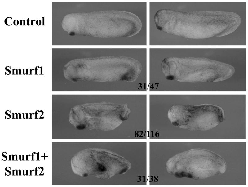
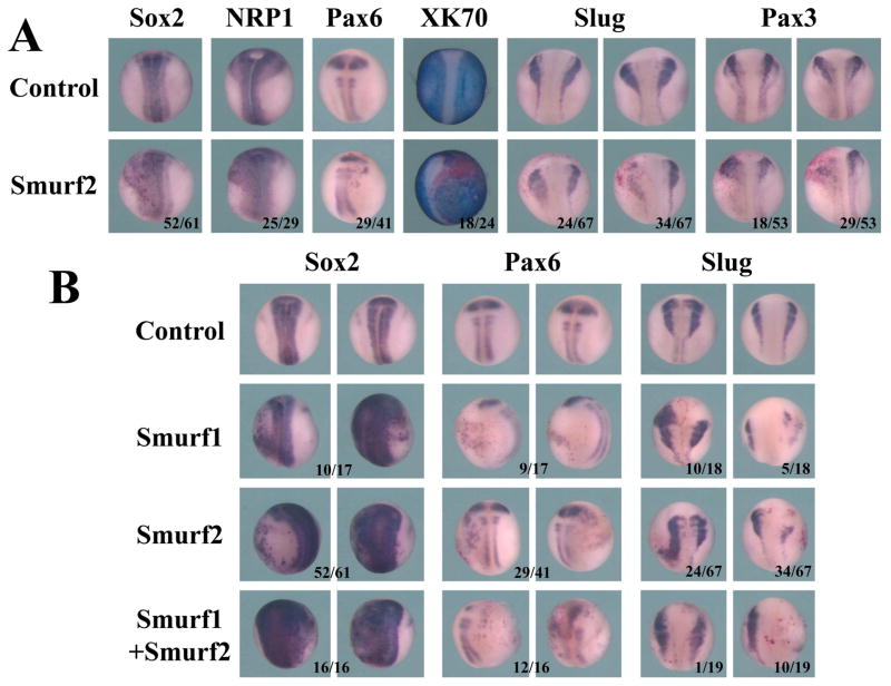
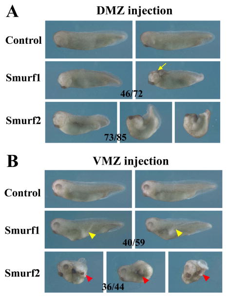
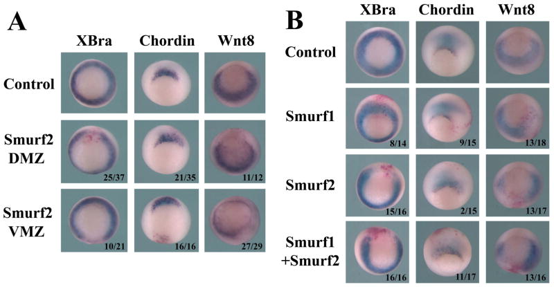
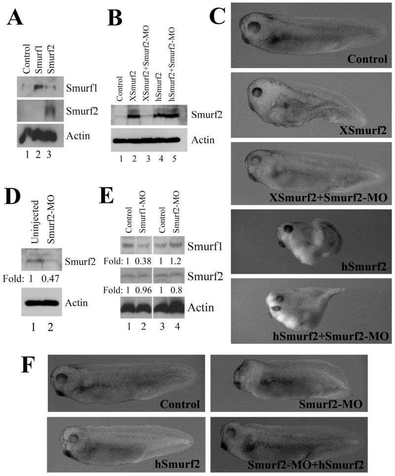
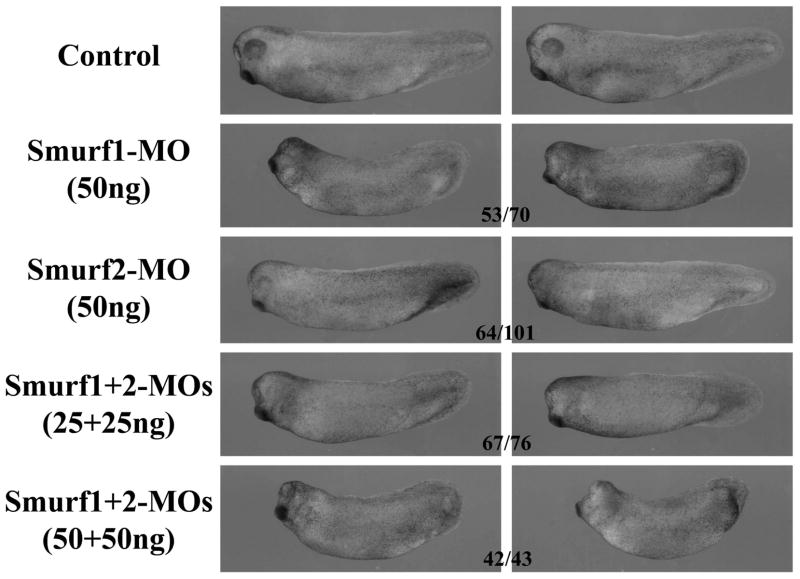
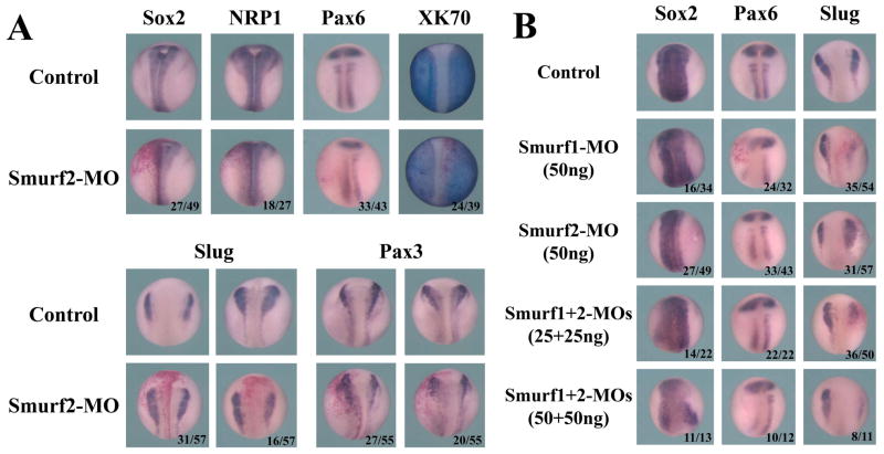
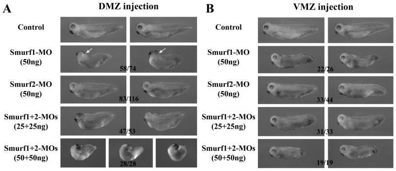
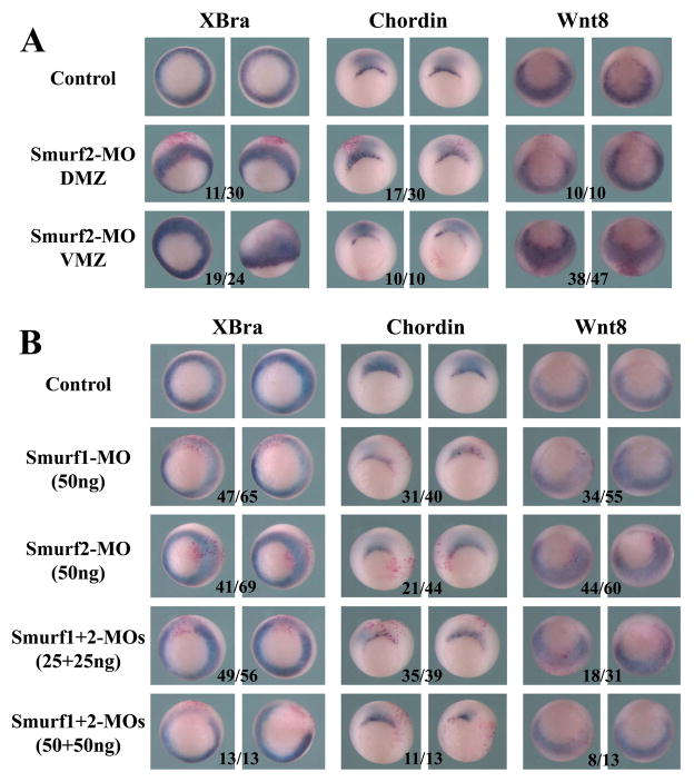
Results: Here we investigate the function of Smurf2 during early Xenopus development. We show that distinctly from Smurf1, overexpression of Smurf2 in presumptive mesoderm interfered with mesoderm induction and caused axial defects, whereas knockdown of Smurf2 with antisense morpholino oligonucleotides resulted in expansion of the mesoderm. These results imply that Smurf2 may modulate nodal-mediated mesodermal induction. Consistently, ventral expression of Smurf2 induced a partial secondary axis with head structures. In the ectoderm, Smurf2 resembled Smurf1 in controlling neural and epidermal marker expression and influencing head formation. Smurf1, but not Smurf2, additionally affected neural tube closure. Interestingly, both Smurfs could enhance as well as repress neural crest markers, implying that they modulate their targets dynamically during neural plate border specification.

Conclusion: Our data demonstrate that Smurf1 and Smurf2 have overlapping and distinct functionalities during early frog embryogenesis; collectively, they regulate ectodermal and mesodermal induction and patterning to ensure normal development of Xenopus embryos.


Figures

Figure 1
Figure 1. Expression pattern of Smurf2 during early Xenopus development
Whole mount in situ hybridization shows that Smurf2 transcripts are uniformly localized to the ectodermal and mesodermal regions in blastula (panels a and b, stage 9 embryos with animal and vegetal views, respectively) and early gastrula (c, stage 10 vegetal view with the arrow pointing to the blastopore lip) embryos. At mid- to late gastrula stages, its expression is enhanced in the dorsal region (panels d and e, stage 11 and 12 embryos, posterior view with dorsal side on top; and panel f, stage 12 embryo lateral view; D: dorsal; V, ventral). During neurulation, Smurf2 is seen in the neural plate and the migration neural crest cells (panels g and h, dorsal and anterior views respectively; NC, neural crest). At the tailbud to tadpole stages, Smurf2 transcripts are detected in the neural tissues, the olfactory and otic vesicles, eyes, migrating neural crest, notochord, and tail (panels i to l, lateral view with the head on the left side; Br: brain; Nt: notochord; Ol, Olfactory placode; Ot: otic vesicle).
Figure 2
Figure 2. Overexpression of Smurfs in the ectoderm induces enlarged or ectopic cement gland and enlarged head
RNAs encoding Smurf1 or 2 were injected into the animal region of early frog embryos and the resulting tailbud embryos are shown here. The numbers indicate the embryos displaying the phenotype.
Figure 3
Figure 3. Elevation of Smurf levels affects neural and neural crest development
A) Increased levels of Smurf2 led to expansion or ectopic expression of the neural genes Sox2, NRP1 and Pax6 and reduction of the epidermal marker XK70, but could both up- and down-regulate the neural plate border genes Slug and Pax3. B) Smurf1 and Smurf2 function similarly to control neural and neural crest development; and together they facilitated expansion of the neural genes but reduction of the neural crest marker Slug. The numbers of the embryos displaying the shown changes in marker expression are indicated in the figure.
Figure 4
Figure 4. Smurf1 and Smurf2 play different roles in mesodermal development
A) Increased Smurf1 expression in dorsal mesoderm results in dorsal-anteriorization of the embryos (46/72 embryos), which often also show anterior neural tube closure defects (in 18 embryos; arrow points to the open brain). In contrast, increased Smurf2 expression in the dorsal mesoderm leads to disruption of axial structures, with embryos frequently displaying gastrulation defects (73/85 embryos with gastrulation defects; among these, 32 embryos had split dorsal structures characterized as the open back phenotype). B) Ectopic expression of Smurf1 in the VMZ induces a partial secondary dorsal axis with the trunk tissues (40/59 embryos; yellow arrowheads); whereas ectopic expression of Smurf2 in the VMZ results in induction of a partial secondary axis that often contains cement gland and hatching gland (36/44 embryos with an ectopic axis, and 25 of these contained cement gland; red arrowheads).
Figure 5
Figure 5. Smurf1 and Smurf2 differentially regulate mesodermal markers at gastrula stages
A) Overexpression of Smurf2 in either DMZ or VMZ disrupts expression of the pan-mesodermal marker Brachyury (XBra). Dorsal expression of Smurf2 slightly reduces the dorsal mesoderm marker chordin, whereas ventral expression of Smurf2 inhibits the ventral-lateral mesodermal marker wnt8. B) Unlike Smurf2, Smurf1 does not block, but instead mildly expands, the expression of XBra or chordin; though like Smurf2, it also inhibits wnt8 expression. Co-expression of Smurf1 and 2 leads to reduction of all three mesodermal markers. The numbers indicate the embryos displaying the shown changes in marker expression.
Figure 6
Figure 6. Smurf2 antisense morpholino oligonucleotides (Smurf2-MO) acts specifically to block translation and function of Xenopus Smurf2
A) Smurf2 antibody recognizes Xenopus Smurf2, but not Smurf1. B) Smurf2-MO inhibits translation from the RNA encoding Xenopus Smurf2 (XSmurf2), but has no effect on translation of human Smurf2 (hSmurf2). C) Smurf2-MO blocks induction of a partial secondary axis by XSmurf2, but not by hSmurf2. D) Smurf2-MO reduces endogenous Smurf2 protein levels at the gastrula stages. E) Smurf1-MO does not regulate the protein level of Smurf2. F) Ectodermal expression of Smurf2-MO induces head defects, which are partially rescued by low doses of co-expressed hSmurf2.
Figure 7
Figure 7. Smurf1 and Smurf2 are both required for normal head development in early Xenopus embryos
Knockdown of either Smurf led to malformation of the head in the resulting tadpoles, with Smurf1 morphants displaying a more severe phenotype. Depletion of both Smurfs exacerbated the head defects, suggesting that the two Smurfs have redundant activities in regulation of head development. The numbers indicate the embryos showing the phenotype.
Figure 8
Figure 8. Smurfs regulate ectodermal marker expression
A) Knockdown of Smurf2 slightly reduced the neural marker expression and expanded the epidermal marker XK70, but the neural plate border genes Slug and Pax3 could be both up- and down-regulated in the morphants. B) Knockdown of Smurf1 resulted in slightly reduced Sox2 in a wider domain and decreased Pax6 expression; Slug was also predominantly reduced (10/54 and 35/54 embryos with enhanced and decreased Slug expression, respectively). Depletion of both Smurf1 and Smurf2 caused reduction and disorganization of the Sox2 domain, and decreased expression of both Pax6 and Slug (8/50 and 36/50 embryos, respectively, with enhanced and reduced Slug in 25ng combination of both MOs, and no embryos showed higher expression of Slug in double morphants with 50ng combination of the MOs).
Figure 9
Figure 9. Smurf1 and Smurf2 control mesendodermal development in early Xenopus embryos
A) Knockdown of Smurf1 in dorsal tissues resulted in reduction of axial structures and malformation of the head, which was often accompanied by a delay or failure in neural tube closure (32 out of the 58 embryos with the shown phenotype had neural tube closure defects, arrow). In contrast, knockdown of Smurf2 led to enlargement of the endodermal mass in the embryos. When both Smurfs were knocked down, the embryos developed with severe axial defects, microcephaly, and failure in gastrulation. B) Knockdown of either Smurf1 or Smurf2 in the VMZ region induced tail truncation, though the phenotype was more pronounced in the Smurf1 morphants. Double knockdown of both Smurfs aggravated the tail defects. The numbers indicate the embryos showing the depicted phenotype.
Figure 10
Figure 10. Smurf1 and Smurf2 differentially regulate mesodermal marker expression
A) Knockdown of Smurf2 in the marginal zone region caused expansion of the pan-mesodermal marker XBra towards the ectodermal region. Dorsal knockdown of Smurf2 reduced expression of chordin, whereas ventral knockdown of Smurf2 led to expansion of wnt8 into the ectodermal domain. B) Unlike Smurf2 knockdown, Smurf1 knockdown reduced the expression of both XBra and chordin, and double knockdown of both Smurfs resulted in more efficient reduction of these markers. The numbers indicate the embryos displaying the shown changes of marker expression.

Similar articles

Cited by

References

    1. Alexandrova EM, Thomsen GH. Smurf1 regulates neural patterning and folding in Xenopus embryos by antagonizing the BMP/Smad1 pathway. Dev Biol. 2006;299:398–410. - PMC - PubMed
    1. Amaravadi LS, Neff AW, Sleeman JP, Smith RC. Autonomous neural axis formation by ectopic expression of the protooncogene c-ski. Dev Biol. 1997;192:392–404. - PubMed
    1. Aybar MJ, Mayor R. Early induction of neural crest cells: lessons learned from frog, fish and chick. Curr Opin Genet Dev. 2002;12:452–458. - PubMed
    1. Baker CV, Bronner-Fraser M. The origins of the neural crest. Part I: embryonic induction. Mech Dev. 1997;69:3–11. - PubMed
    1. Barembaum M, Bronner-Fraser M. Early steps in neural crest specification. Semin Cell Dev Biol. 2005;16:642–646. - PubMed

Publication types

MeSH terms

LinkOut - more resources