. 2012 Jul 20;287(30):25395-406.
doi: 10.1074/jbc.M112.370130. Epub 2012 Jun 6.

A potential neuroprotective role of apolipoprotein E-containing lipoproteins through low density lipoprotein receptor-related protein 1 in normal tension glaucoma

Affiliations

A potential neuroprotective role of apolipoprotein E-containing lipoproteins through low density lipoprotein receptor-related protein 1 in normal tension glaucoma

Hideki Hayashi et al. J Biol Chem. .

Abstract

Glaucoma is an optic neuropathy and the second major cause of blindness worldwide next to cataracts. The protection from retinal ganglion cell (RGC) loss, one of the main characteristics of glaucoma, would be a straightforward treatment for this disorder. However, the clinical application of neuroprotection has not, so far, been successful. Here, we report that apolipoprotein E-containing lipoproteins (E-LPs) protect primary cultured RGCs from Ca(2+)-dependent, and mitochondrion-mediated, apoptosis induced by glutamate. Binding of E-LPs to the low density lipoprotein receptor-related protein 1 recruited the N-methyl-d-aspartate receptor, blocked intracellular Ca(2+) elevation, and inactivated glycogen synthase kinase 3β, thereby inhibiting apoptosis. When compared with contralateral eyes treated with phosphate-buffered saline, intravitreal administration of E-LPs protected against RGC loss in glutamate aspartate transporter-deficient mice, a model of normal tension glaucoma that causes glaucomatous optic neuropathy without elevation of intraocular pressure. Although the presence of α2-macroglobulin, another ligand of the low density lipoprotein receptor-related protein 1, interfered with the neuroprotective effect of E-LPs against glutamate-induced neurotoxicity, the addition of E-LPs overcame the inhibitory effect of α2-macroglobulin. These findings may provide a potential therapeutic strategy for normal tension glaucoma by an LRP1-mediated pathway.


Figures

FIGURE 1.
FIGURE 1.
Glutamate with glycine induces apoptosis in RGCs. A, fragmented or shrunken nuclei in RGCs were detected by Hoechst staining 24 h after control (C; HBSS) or treatment with glutamate alone (Glu(); 300 μm glutamate), glycine alone (Gly(); 10 μm glycine), or glutamate + glycine (Glu; 300 μm glutamate + 10 μm glycine). Data are means ± S.E. from 4 independent experiments. *, p < 0.001 for control versus Glu. B, fragmented or shrunken nuclei were detected by Hoechst staining 24 h after control (HBSS) or glutamate + glycine treatment (Glu; 300 μm glutamate plus 10 μm glycine) with 0, 15 (one 15-min wash), 30 (two 15-min washes), or 45 min (three 15-min washes) of washing by HBSS. C, fluorescence images of RGCs stained with annexin V-EGFP, propidium iodide (PI), and Hoechst 12 h after control (HBSS) or Glu treatment (300 μm glutamate + 10 μm glycine). Scale bar, 20 μm. D, RGCs stained with 2 nm MitoTracker Red (Mito) were immunostained with anti-cytochrome c (Cyto C) 12 h after control or Glu treatment. Scale bar, 20 μm.
FIGURE 2.
FIGURE 2.
Contributors to glutamate-induced neurotoxicity in RGCs. Fragmented or shrunken nuclei were detected by Hoechst staining 24 h after control (C; HBSS) or glutamate treatment (Glu; 300 μm glutamate + 10 μm glycine). A, Glu was incubated with the cells with or without Ca2+. B–F, RGCs were treated with 10 μm MK801 (inhibitor of the NMDA receptor) (B), 1 μm N-acetyl-leucine leucine norleucinal (ALLN) (calpain inhibitor) (C), 1 μm FK506 (calcineurin inhibitor) (D), 200 μm Bax-inhibitory peptide V5 (BIP-V5; inhibitor of Bax) (E), 20 μm Z-VAD-fmk (Z-VAD; caspase inhibitor) (F), or dimethyl sulfoxide (DM) when Glu was added. In B–F, *, p < 0.001 for Glu versus Glu+inhibitor. Data are means ± S.E. from 4–6 independent experiments.
FIGURE 3.
FIGURE 3.
Lipoproteins prevent apoptosis induced by glutamate. Fragmented or shrunken nuclei were detected by Hoechst staining 24 h after control (C; HBSS) or glutamate treatment (Glu; 300 μm glutamate + 10 μm glycine). A, immunoblot for apoE in lipoproteins (2 μg of cholesterol/ml) isolated from glia-conditioned medium (GLP, glia-derived E-LPs), HDL (2 μg of cholesterol/ml) isolated from rat serum and reconstituted human apoE-containing lipoproteins (100 ng of protein/ml) (E-LP). RGCs were incubated for 15 min with GLP, HDL, or E-LP, and then Glu was added as indicated. *, p < 0.005 for Glu versus Glu+GLP, Glu+HDL or Glu+E-LP. B, dose-dependent protection of E-LP against glutamate neurotoxicity. RGCs were incubated for 15 min with the indicated concentrations of E-LP, and then Glu was added. The percentage of protection by E-LPs was calculated such that control was 100% and glutamate treatment without E-LP was 0% of protection. C, RGCs were incubated with lipid-free apoE (100 ng of protein/ml), cholesterol (chol) (11 ng/ml), phosphatidylcholine+cholesterol (PC+chol) liposomes (11 ng of cholesterol/ml), PC+apoE lipoproteins (100 ng of protein/ml), E-LP containing apoE, cholesterol and phosphatidylcholine (100 ng of protein and 11 ng of cholesterol/ml), HDL (2 μg of cholesterol/ml) from mouse plasma (Ms HDL), or HDL (2 μg of cholesterol/ml) from apoE-deficient mouse plasma (E−/− HDL), and then Glu was added as indicated. * and **, p < 0.001 and 0.0001, respectively, for Glu versus Glu+lipoproteins. D, RGCs were incubated with reconstituted human apoE3- (E3-LP) or apoE4-containing lipoproteins (E4-LP) for 15 min, and then Glu was added as indicated. *, p < 0.001 for Glu versus Glu+E3-LP or Glu+E4-LP.
FIGURE 4.
FIGURE 4.
E-LPs inhibit the elevation of intracellular Ca2+ by interaction of LRP1 with the NMDA receptor. A, RGCs were labeled with Fluo-8 acetoxymethyl ester for 30 min, and then Glu (300 μm glutamate + 10 μm glycine) was added ± E-LP (100 ng of protein/ml). Fluorescence ratio images are displayed in pseudocolor as indicated by the color bar at the bottom. Pseudocolor represents changes in fluorescence ratios between 0 (blue) and 2 (red) corresponding to 1 (green), which is defined as the basal fluorescence intensity before Glu stimulation. Left and right panels show ratio images with Glu and Glu+E-LP, respectively. Data are from one experiment representative of 8 experiments with similar results. Scale bar, 80 μm. B and C, changes in Fluo-8 fluorescence are expressed as ΔF/F0, where F0 is basal fluorescence intensity before Glu stimulation. RGCs were incubated with 100 ng of protein/ml E-LP, 10 μm MK801, 100 ng of protein/ml E-LP+10 μm MK801, 100 ng of protein/ml E-LP+10 μg/ml anti-LRP1 antibody, or 100 ng of protein/ml E-LP+10 μg/ml IgG for 15 min, and then Glu was added as indicated. Data are means ± S.E. from 6–8 independent experiments. D, fragmented or shrunken nuclei were detected by Hoechst staining 24 h after control (C; HBSS) or Glu treatment. E-LP (100 ng of protein/ml), E-LP+anti-LRP1 (10 μg/ml), or E-LP+IgG (10 μg/ml) was added to RGCs for 15 min, and then Glu was added. *, p < 0.01 for Glu+E-LP versus Glu+E-LP+anti-LRP1. Data are means ± S.E. from 4 independent experiments. E and F, LRP1 (E) or NMDA receptor subunit NR2B (F) was immunoprecipitated from lysates of RGCs treated ± E-LP for 15 min. Immunoprecipitates (Pellet) and supernatant were probed with antibodies raised against LRP1, NR2B, or NR2A. Data are from one experiment representative of 3 experiments with similar results.
FIGURE 5.
FIGURE 5.
Phospholipase C, protein kinase Cδ, and GSK3β contribute to the protective effect of E-LP against glutamate neurotoxicity. A, fragmented or shrunken nuclei were detected by Hoechst staining 24 h after control (C; HBSS) or glutamate (Glu; 300 μm glutamate + 10 μm glycine) treatment. RGCs were incubated with 100 ng of protein/ml E-LP or E-LP+U (U, 5 μm U73122, phospholipase C inhibitor) for 15 min, and then Glu was added. Data are means ± S.E. from 5 independent experiments. *, p < 0.005 for Glu+E-LP versus Glu+E-LP+U. B, knockdown of protein kinase Cδ (PKCδ) in RGCs was induced by PKCδ siRNA. RGCs were incubated with 300 nm negative control (NC) or PKCδ siRNA for 6 days, and then PKCδ was detected by immunoblotting. β-Actin was used as loading control. Fragmented or shrunken nuclei were detected by Hoechst staining 24 h after control, Glu, or Glu+E-LP treatment with knockdown by negative control or PKCδ siRNA. Data are means ± S.E. from 5 independent experiments. *, p < 0.05 for Glu+E-LP+NC versus Glu+E-LP+PKCδ. C, RGCs were incubated for 16 h after control, Glu, or Glu+E-LP treatment. RGC proteins were immunoblotted with antibodies raised against GSK3β phosphorylated at Ser-9 (p-GSK3β) or total GSK3β (GSK3β). Quantification of Ser9 phosphorylation of GSK3β is shown from 4 independent experiments. *, p < 0.001 for Glu versus Glu+E-LP.
FIGURE 6.
FIGURE 6.
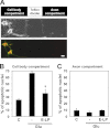
Apoptosis is induced by glutamate in cell body, but not distal axon, compartments. A, upper panel, phase contrast image of RGCs in one track shows that cell bodies are localized to the cell body compartment and that distal axons are present in the right-hand axon compartment. Bottom panel, RGCs were stained with anti-LRP1 (green) and anti-NR2B (red) antibodies. Scale bar, 50 μm. B and C, fragmented or shrunken nuclei were detected by Hoechst staining 24 h after control (C; HBSS) or glutamate (Glu; 300 μm glutamate + 10 μm glycine) treatment of cell bodies (B) or distal axons (C). Cell bodies and axons of RGCs were incubated with 100 ng of protein/ml E-LP for 15 min, and then Glu was added to cell bodies (B) and distal axons (C). *, p < 0.005 for Glu versus Glu+E-LP. B and C, data are means ± S.E. from 4 independent experiments.
FIGURE 7.
FIGURE 7.
E-LPs restore RGC survival in Glast+/− and Glast−/− mice. A and B, retinae from 3-week-old (3w) and 6-week-old (6w) Glast−/− mice injected with 1 μl of PBS or 1 μl of 1.5 μg of protein/ml E-LPs were subjected to immunoblotting with anti-Brn-3a and anti-β-actin antibodies (A) or antibodies raised against GSK3β phosphorylated at Ser-9 (p-GSK3β) and total GSK3β (GSK3β) (B). A and B, quantification of Brn-3a relative to β-actin (A) and phosphorylation at Ser-9 of GSK3β relative to total GSK3β (B) from 5 and 6 independent experiments, respectively. *, p < 0.05 for PBS versus E-LP in 6-week-old Glast−/− mice. C, hematoxylin and eosin staining of a retinal section from Glast+/− and Glast−/− mice (3 or 6 weeks old) injected with 1 μl of PBS, 1 μl of 1.5 μg of protein/ml E-LPs, or 30 μg of cholesterol/ml HDL. Arrowhead indicates ganglion cell layer of retina (GCL). Scale bar represents 40 μm. Data are from one retinal section representative of 8 retinae with similar results. D, quantification of number of RGCs in 6-week-old wild type (+/+), 3- and 6-week-old Glast+/− (+/−), and Glast−/− (−/−) mice injected without (NT: no treatment) or with 1 μl of PBS, 1 μl of 1.5 μg of protein/ml E-LPs, or 1 μl of 30 μg of cholesterol/ml HDL. Data are from 8 independent experiments. *, p < 0.05 for PBS versus E-LP or HDL in 6-week-old Glast−/− mice. #, p < 0.05 for PBS versus E-LP or HDL in 6-week-old Glast+/− mice. GC layer, ganglion cell layer.
FIGURE 8.
FIGURE 8.
Inhibitory effect of α2-macroglobulin is overcome by E-LPs. A and B, retinae and vitreous humor from 3-week-old (3w) and 6-week-old (6w) Glast+/+ and Glast−/− mice were immunoblotted with antibodies raised against apoE, LRP1, β-actin, α2-macroglobulin (a2M),and albumin. Arrowhead indicates a2M. A, quantification of apoE relative to β-actin from 4 independent experiments. *, p < 0.05 for 3-week-old Glast+/+ versus 3-week-old Glast−/− retina. B, quantification of apoE and a2M relative to albumin from 3 independent experiments. *, p < 0.05 for 3-week-old Glast+/+ versus 3-week-old Glast−/− vitreous humor. #, p < 0.05 for 6-week-old Glast+/+ versus 6-week-old Glast−/− vitreous humor. C, fragmented or shrunken nuclei in RGCs were detected by Hoechst staining 24 h after control (C; HBSS), glutamate (Glu; 300 μm glutamate + 10 μm glycine), or Glu+100 ng of protein/ml E-LP treatment ± 100 nm a2M. *, p < 0.05 for Glu+E-LP versus Glu+E-LP+a2M. D, RGCs were incubated with 100 nm a2M and E-LP (100–3000 ng of protein/ml) for 15 min, and then Glu was added. C and D, data are means ± S.E. from 4 independent experiments. * and **, p < 0.05 and 0.005, respectively, for Glu versus Glu +a2M+E-LP.
FIGURE 9.
FIGURE 9.
Proposed protective pathway by E-LP against glutamate-induced apoptosis in RGCs. E-LPs protect RGCs from glutamate-induced apoptosis via an LRP1-mediated signaling pathway. Glutamate (Glu) induces the elevation of intracellular Ca2+ and activates calpain and calcineurin (CaN). Calcineurin dephosphorylates phospho (P)-Bax and leads to mitochondrion (cytochrome c; Cyt c)-mediated and caspase (Casp)-dependent apoptosis. When E-LPs bind to LRP1, the formation of an LRP1-NMDA receptor (NMDAR) complex is promoted and attenuates the elevation of intracellular Ca2+ caused by glutamate. Thus, RGCs are protected from neurodegeneration. In addition, binding of E-LPs to LRP1 activates phospholipase Cγ1 (PLCγ1), and PKCδ then increases the phosphorylation (P) of the proapoptotic kinase GSK3β, thereby inactivating GSK3β. α2-Macroglobulin (a2M), directly or indirectly, interrupts the protective effect of E-LPs. PIP2, phosphatidylinositol 4,5-bisphosphate; IP3, inositol 1,4,5-trisphosphate; DAG, diacylglycerol.

References

    1. Resnikoff S., Pascolini D., Etya'ale D., Kocur I., Pararajasegaram R., Pokharel G. P., Mariotti S. P. (2004) Global data on visual impairment in the year 2002. Bull. World Health Organ 82, 844–851 - PMC - PubMed
    1. Quigley H. A. (2011) Glaucoma. Lancet 377, 1367–1377 - PubMed
    1. Iwase A., Suzuki Y., Araie M., Yamamoto T., Abe H., Shirato S., Kuwayama Y., Mishima H. K., Shimizu H., Tomita G., Inoue Y., Kitazawa Y. (2004) The prevalence of primary open-angle glaucoma in Japanese: the Tajimi Study. Ophthalmology 111, 1641–1648 - PubMed
    1. Pekmezci M., Vo B., Lim A. K., Hirabayashi D. R., Tanaka G. H., Weinreb R. N., Lin S. C. (2009) The characteristics of glaucoma in Japanese Americans. Arch. Ophthalmol. 127, 167–171 - PubMed
    1. Mansberger S. L., Romero F. C., Smith N. H., Johnson C. A., Cioffi G. A., Edmunds B., Choi D., Becker T. M. (2005) Causes of visual impairment and common eye problems in Northwest American Indians and Alaska Natives. Am. J. Public Health 95, 881–886 - PMC - PubMed

Publication types

MeSH terms