Review
. 2012 Nov;85 Spec No 1(Spec Iss 1):S41-53.
doi: 10.1259/bjr/30029741. Epub 2012 Jun 6.

Focal testicular lesions: colour Doppler ultrasound, contrast-enhanced ultrasound and tissue elastography as adjuvants to the diagnosis

Affiliations
Review

Focal testicular lesions: colour Doppler ultrasound, contrast-enhanced ultrasound and tissue elastography as adjuvants to the diagnosis

D Y Huang et al. Br J Radiol. 2012 Nov.

Abstract

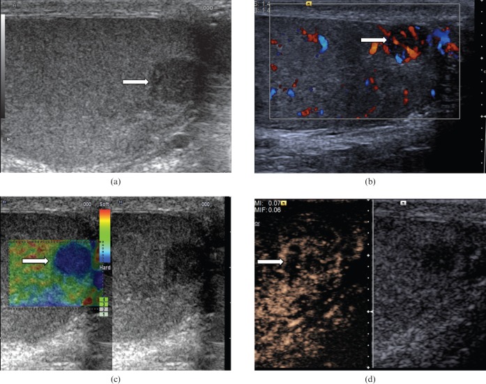
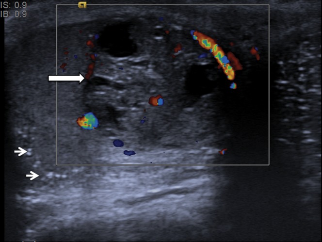
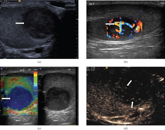
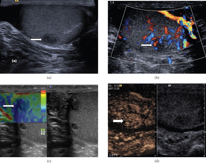
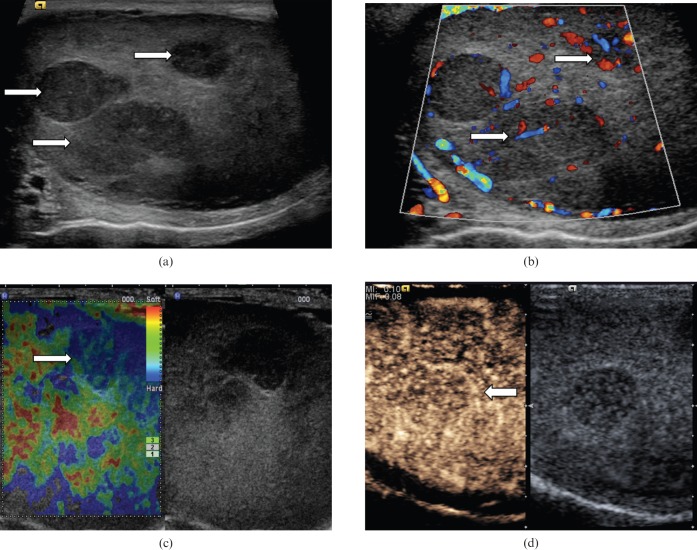
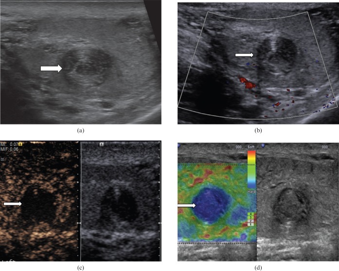
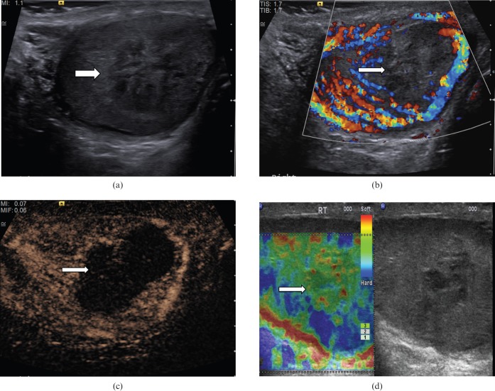
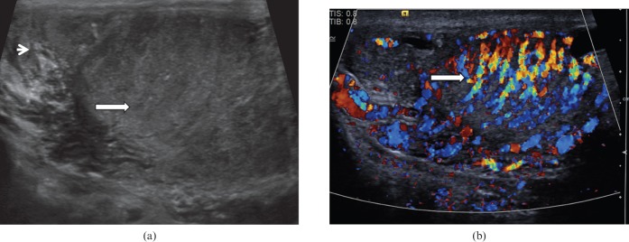
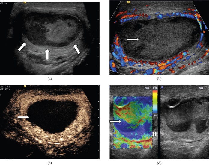
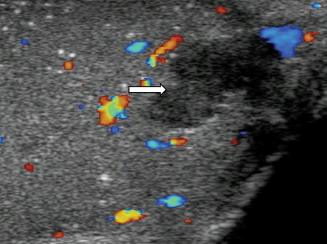
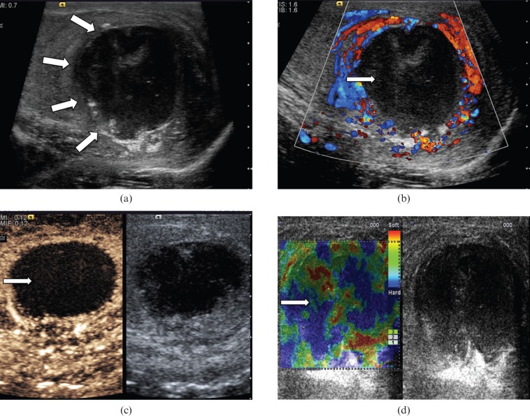
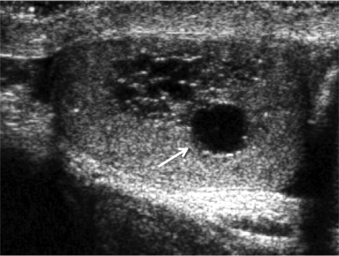
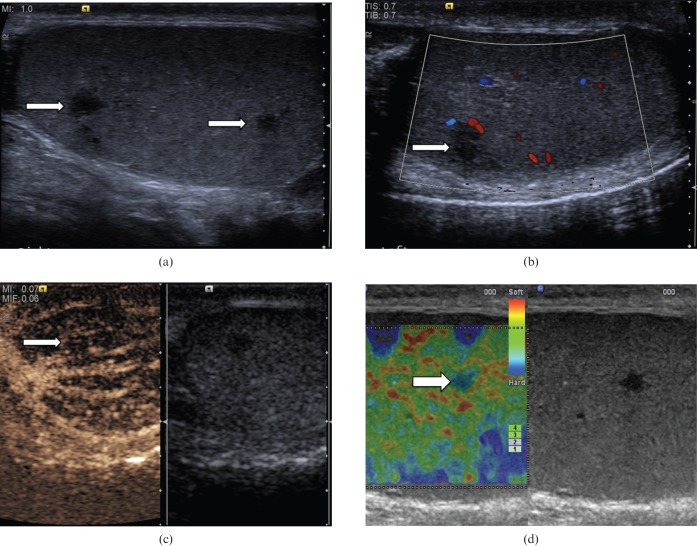
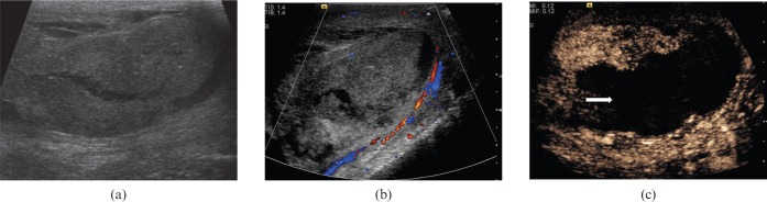
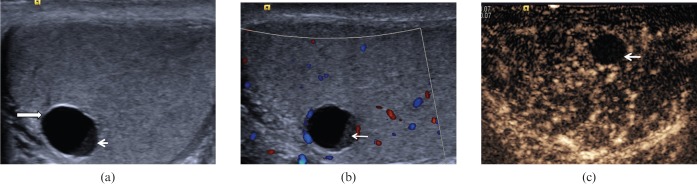
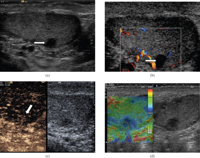
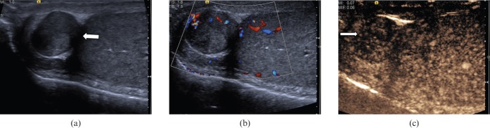
The aim of this review is to illustrate the potential of different and newer ultrasound techniques beyond conventional B-mode imaging, including colour Doppler ultrasound, contrast-enhanced ultrasound (CEUS) and tissue elastography, in the characterisation of both benign and malignant intratesticular lesions. Normally, testicular malignancies, either primary or secondary, demonstrate an increase in colour Doppler signal. However, there is a diversity of benign testicular lesions that may mimic testicular malignancies. The use of CEUS improves characterisation of testicular lesions, and confirms lack of vascularity in benign abnormalities such as epidermoid cysts, infarctions, abscesses and changes following trauma. Tissue elastography allows further evaluation of the cellular consistency of the abnormality. Familiarity with the appearances seen with these ultrasound techniques in both benign and malignant abnormalities should aid in improving confidence in arriving at the correct diagnosis.


Figures

Figure 1
Figure 1
Seminoma. (a) B-mode ultrasound demonstrates a small focal lesion (arrow) measuring 6 mm, with uniform low reflectivity. (b) Colour Doppler ultrasound demonstrates internal vascularity within the small tumour (arrow). (c) The lesion appears “hard” on tissue elastography (depicted by the blue area, arrow). (d) Contrast-enhanced ultrasound demonstrates early enhancement and rapid washout with loss of the normal vascular pattern.
Figure 2
Figure 2
Mixed germ cell tumour. A focal lesion with heterogeneous reflectivity and cystic components (long arrow). Colour Doppler demonstrates distortion of the normal vascular pattern by the lesion. Note is also made of background testicular microlithiasis (short arrows).
Figure 3
Figure 3
Embryonal cell tumour. (a) B-mode ultrasound demonstrates a focal lesion with a slightly heterogeneous reflectivity (arrow). (b) Colour Doppler ultrasound demonstrates loss of normal parenchymal vascular pattern, replaced by an abnormal vascularity (the “criss-cross” vascular pattern; arrow). Colour Doppler flow is demonstrated in the large vessels only. (c) Tissue elastography demonstrates a “blue” lesion, therefore clearly a “hard” lesion (arrow). (d) On contrast-enhanced ultrasound, particulate movement of contrast (arrows) is seen throughout the lesion in a haphazard pattern, confirming the vascularity is present within all components of the lesion.
Figure 4
Figure 4
Leydig cell tumour. (a) B-mode ultrasound demonstrates a small hypoechoic lesion (arrow). (b) Colour Doppler ultrasound demonstrates increased vascularity within the lesion (arrow). (c) Tissue elastography demonstrates a small distinct “hard” lesion (mixed blue/green area, arrow). (d) With contrast-enhanced ultrasound the lesion (arrow) demonstrates early enhancement, a characteristic that has the potential to differentiate these lesions from other malignant tumours.
Figure 5
Figure 5
Prostatic metastasis. (a) B-mode ultrasound demonstrates multifocal hypoechoeic lesions (arrows). (b) Colour Doppler ultrasound demonstrates internal vascularity within the lesions (arrows). (c) The lesions appear “hard” on elastography (blue/green area, arrow). (d) Enhancement is noted within the lesion, confirming internal vascularity and peripheral contrast enhancement (arrow).
Figure 6
Figure 6
Epidermoid cyst. (a) B-mode ultrasound demonstrates a well-circumscribed, solid, mixed-reflectivity lesion with high-reflectivity “onion-skin” rims (arrow). (b) Colour Doppler signal is not observed within the lesion (arrow). (c) Contrast-enhanced ultrasound demonstrates a clear lack of enhancement within the lesion (arrow). (d) Tissue elastography demonstrates a well-demarcated “hard” lesion, blue in colour (arrow).
Figure 7
Figure 7
Segmental testicular infarction. (a) There is a focal mixed-reflectivity area (arrow) in a patient who presented with testicular pain and clinical evidence of epididymo-orchitis. (b) There is no colour Doppler signal within the abnormality, suggesting absence of vascularity (arrow). (c) Contrast-enhanced ultrasound clearly depicts the infarcted areas with absence of enhancement (arrow). (d) Tissue elastography demonstrates that the abnormality is “soft” (red/green on colour scale, arrow) and no focal “hard” lesion is visualised, in contrast to the cases of focal testicular tumours.
Figure 8
Figure 8
Orchitis. (a) Longitudinal ultrasound of the testis demonstrates patchy heterogeneous reflectivity within the testis (long arrow) and enlargement of the epididymis (short arrow). (b) There is marked increase in vascularity within the testis on colour Doppler ultrasound (arrow).
Figure 9
Figure 9
Venous infarction of the testis. (a) A focal testicular abnormality with mixed reflectivity (arrows) is noted on B-mode ultrasound. (b) No colour Doppler signal is seen in the focal testicular abnormality (arrow). (c) Following the administration of microbubble contrast, contrast flow is present in the normal testicular parenchymal, and clearly absent from the infarcted portion of the testis (arrow). (d) Tissue elastography demonstrates no focal “hard” lesions, and the area of abnormality appears “soft” (green on colour scale, arrow).
Figure 10
Figure 10
Intratesticular haematoma. A well-circumscribed (arrow) focal area of low reflectivity with internal echoes is noted in the testis of a patient involved in a motorcycle accident. The lesion demonstrates low reflectivity. Colour Doppler ultrasound confirms absence of vascularity, in keeping with the diagnosis of traumatic intratesticular haematoma. At 4 weeks there was reduction in size of the lesion. Incidental microlithiasis is present.
Figure 11
Figure 11
Testicular abscess. (a) On B-mode ultrasound a focal lesion with low internal echoes (arrows) is seen in a patient with history of resolving epididymo-orchitis. (b) On colour Doppler ultrasound there is increased vascularity at the periphery of the lesion but none within the lesion (arrow). (c) Contrast-enhanced ultrasound image demonstrating increased absence of vascularity in the abscess (arrow) with some rim enhancement. (d) Tissue elastography demonstrates a heterogeneous pattern of firmness but no focal “hard” lesion is demonstrated (arrow).
Figure 12
Figure 12
Rete testis. Localised area of tubular ectasia of the rete testis, with a further testicular cyst (arrow). No soft tissue component or internal colour Doppler signal (not shown) is demonstrated to suggest the presence of an underlying tumour.
Figure 13
Figure 13
Testicular sarcoidosis. (a) B-mode ultrasound demonstrates multiple low-reflectivity focal testicular lesions (arrows) in a patient with a recent clinical diagnosis of sarcoidosis. (b) On the colour Doppler study the focal lesions do not clearly demonstrate vascular flow, but it is difficult to be certain because of the size of the lesions. (c) Contrast-enhanced ultrasound clearly confirms some vascularity within the lesion (arrow). (d) Tissue elastography demonstrates a moderate degree of “hardness” of these lesions (blue on colour scale, arrow).
Figure 14
Figure 14
Post-traumatic testicular devascularisation. (a) On B-mode images demonstrates the testis appears very heterogeneous and appears to be “shattered”. (b) There is no clear evidence of colour Doppler flow to the affected testis. (c) Following administration of microbubble contrast, the testis is seen to be predominantly devascularised. The abnormality is much better demarcated on contrast-enhanced ultrasound (arrow).
Figure 15
Figure 15
Cyst with debris. (a) A 6-mm anechoic lesion (long arrow) is noted in the testicle with a thin clear wall demonstrated. A “fluid-debris” level is noted (short arrow). (b) No internal colour Doppler signal is demonstrated within the debris present in the lower aspect of the cyst (arrow). (c) On contrast-enhanced ultrasound, there is clear absence of enhancement in the debris present in the lower aspect of the cyst (arrow), and a cystic tumour is unlikely.
Figure 16
Figure 16
Post-biopsy scar. (a) On B-mode image there is a 3.1×4.6-mm low-reflectivity lesion (arrow) in the mid-aspect of the testicle, which corresponds to the site of a previous testicular biopsy. (b) No internal colour Doppler vascular signal is demonstrated (arrow). (c) There is some subtle enhancement (arrow) on the contrast-enhanced ultrasound examination. (d) Tissue elastography suggests the area is slightly “harder” than the adjacent normal parenchyma (blue area, strain ratio of 1.33). Follow-up ultrasound 1 year later demonstrated no interval changes in appearances of the lesion.
Figure 17
Figure 17
Extratesticular adenomatoid lesion. (a) A heterogeneous lesion (arrow) measuring 12 mm in diameter is noted within the right epididymal head. (b) Colour Doppler signal is demonstrated within the lesion. (c) Following contrast administration the focal epididymal lesion demonstrates enhancement and early washout (arrow).

Similar articles

Cited by

References

    1. Horstman WG, Melson GL, Middleton WD, Andriole GL. Testicular tumors: findings with color Doppler US. Radiology 1992;185:733–7 - PubMed
    1. Lock G, Schmidt C, Helmich F, Stolle E, Dieckmann KP. Early experience with contrast-enhanced ultrasound in the diagnosis of testicular masses: a feasibility study. Urology 2011;77:1049–53 - PubMed
    1. Hedayati V, Sellars ME, Sharma DM, Sidhu PS. Contrast-enhanced ultrasound in testicular trauma: role in directing debridement and aiding organ salvage. Br J Radiol 2012;85:e65–8 - PMC - PubMed
    1. Shah A, Lung PF, Clarke JL, Sellars ME, Sidhu PS. New ultrasound techniques for imaging of the indeterminate testicular lesion may avoid surgery completely. Clin Radiol 2010;65:496–8 - PubMed
    1. Goddi A, Sacchi A, Magistretti G, Almolla J, Salvador M. Real-time tissue elastography for testicular lesion assessment. Eur Radiol 2012;22:721–30 - PMC - PubMed