

. 2012 Nov;85 Spec No 1(Spec Iss 1):S94-101.
doi: 10.1259/bjr/62473200.
Epub 2012 Jun 6.
Ultrasound of the male anterior urethra
Affiliations
- PMID: 22674713
- PMCID: PMC3746405
- DOI: 10.1259/bjr/62473200
Item in Clipboard
Ultrasound of the male anterior urethra
Br J Radiol.
2012 Nov.
Abstract
Imaging of the anterior male urethra has traditionally been performed by fluoroscopic contrast urethrography. While providing easily interpretable images, this technique has a number of disadvantages associated with it. An alternative approach is to use ultrasound to assess the lumen of the urethra and the periurethral tissues. Here we describe the development of urethral ultrasound and the ascending and descending urethral ultrasound techniques employed in our institution with reference to commonly and uncommonly encountered pathologies. We also identify common pitfalls and how to avoid them.
Figures
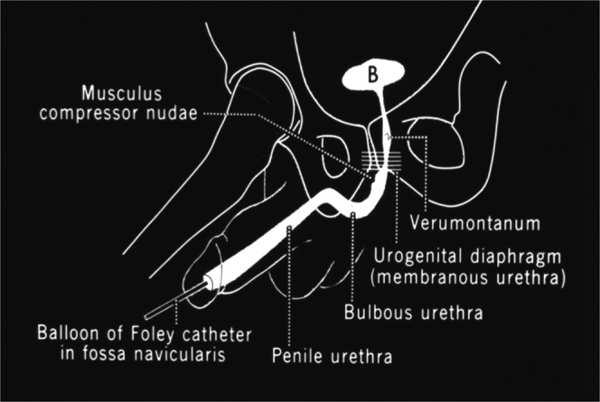
Anatomy and technique of retrograde contrast urethrography [1].
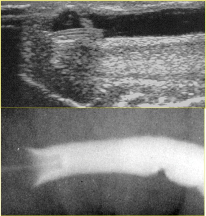
Demonstration of comparative ultrasound (top) and contrast urethrography (bottom) images of the distal urethra.
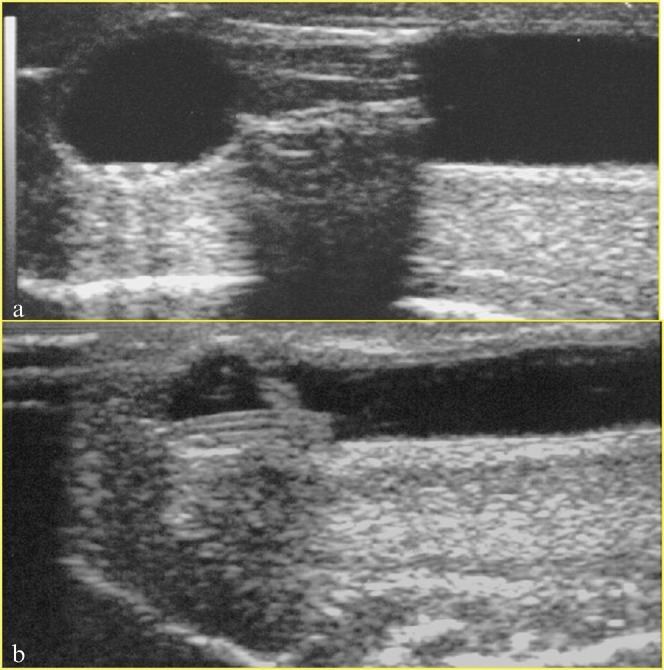
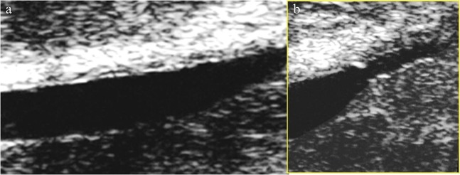
(a) Posterior acoustic shadowing behind the tip of the catheter obscuring the distal urethra. (b) Better demonstration of the distal urethra following trimming of the distal catheter beyond the balloon.
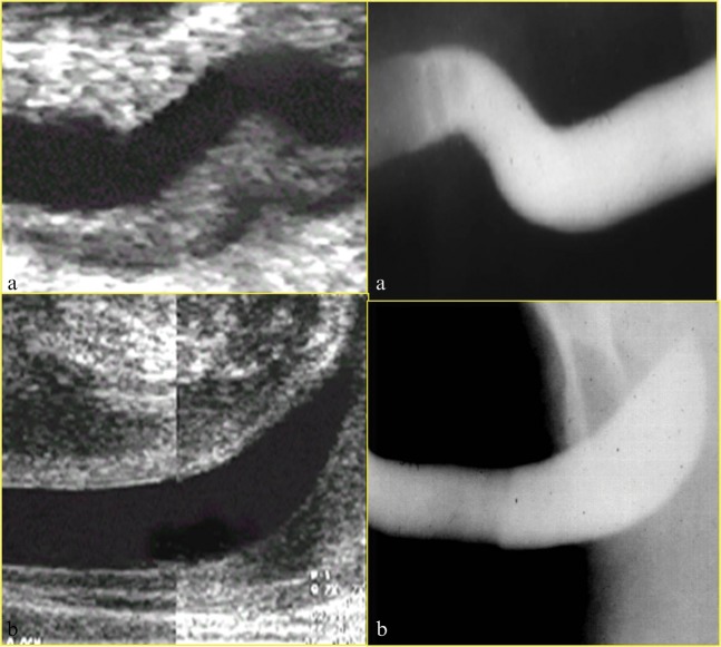
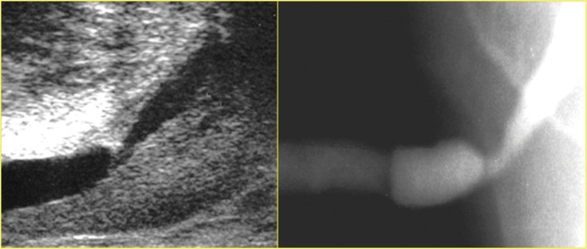
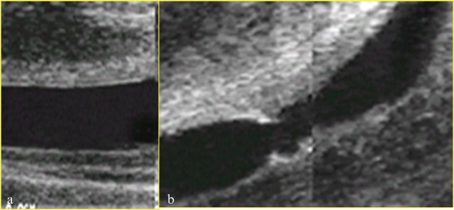
Comparison of normal urethral anatomy as seen on ultrasound (left) and contrast urethrography (right). (a) Normal peno-scrotal junction and (b) normal bulbar urethra.
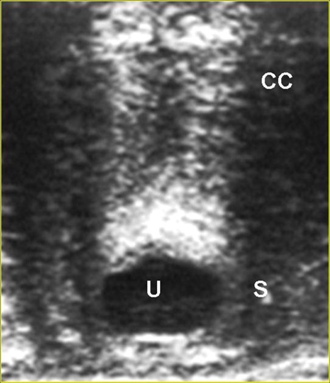
Normal urethra in transverse section. CC, corpus cavernosum; S, spongiosum surrounding urethra; U, urethral lumen.
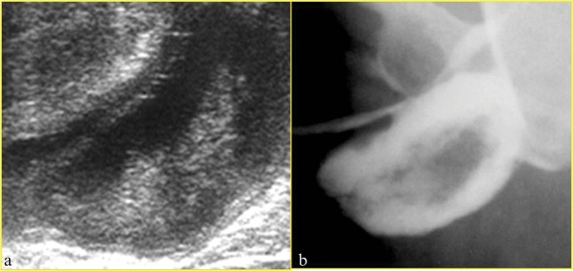
Bulbar stricture on ultrasound (left) and urethrography (right).
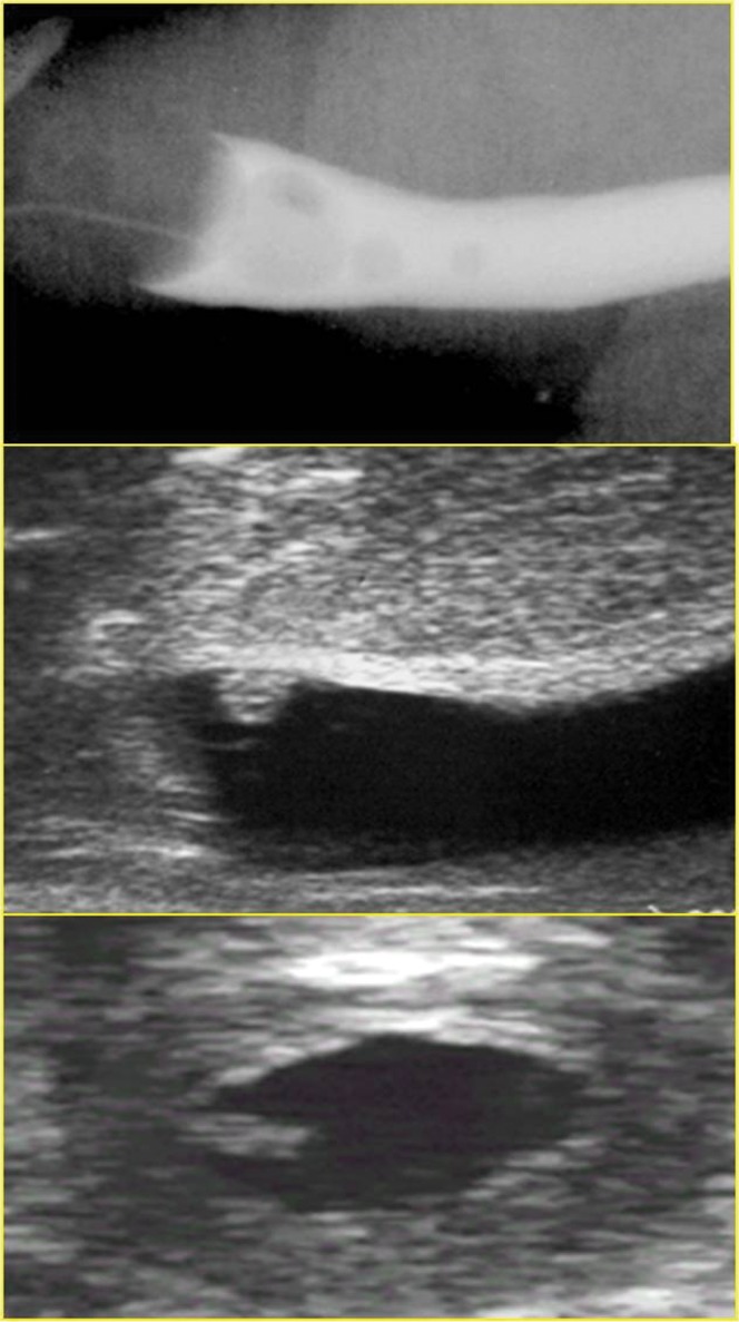
The viral papilloma demonstrated on the ultrasound study (middle and bottom) was overlooked as a bubble on the contrast urethrogram (top).
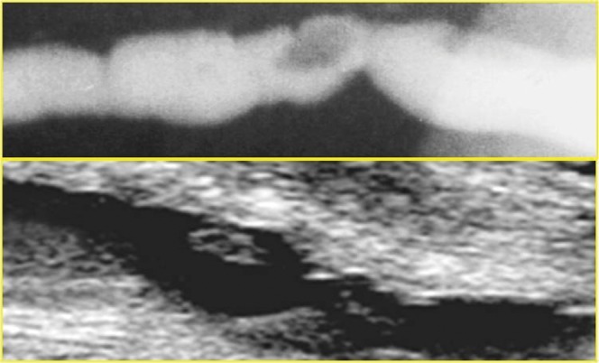
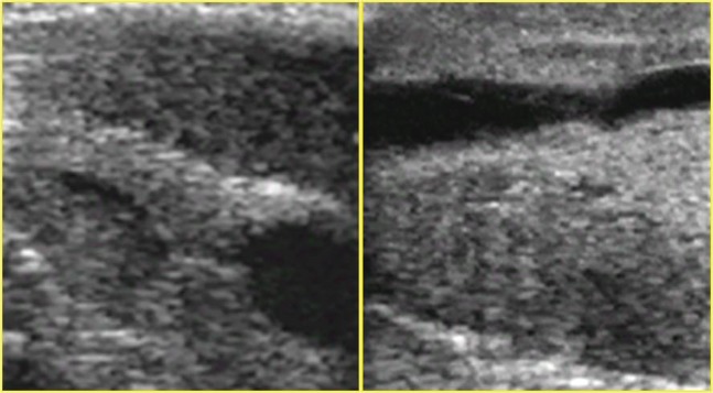
Mucosal tag seen as filling defect on urethrography (top) and ultrasound (bottom).
(a) Urethral diverticulum seen on ultrasound with layering of debris within it and (b) urethrogram correlate.
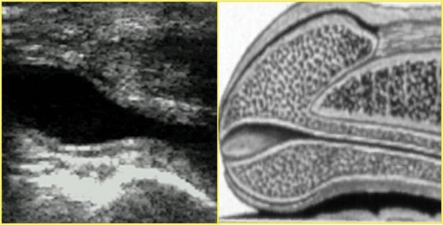
Normal navicular fossa seen during voiding descending urethral ultrasound (left), with diagrammatic representation of navicular fossa (right).
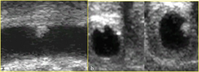
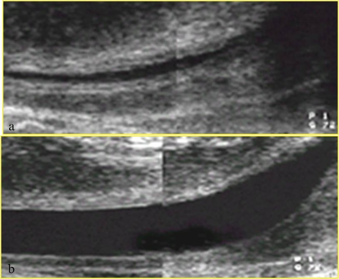
Urethral stricture seen using the descending ultrasound technique in (a) longitudinal and (b) transverse planes.
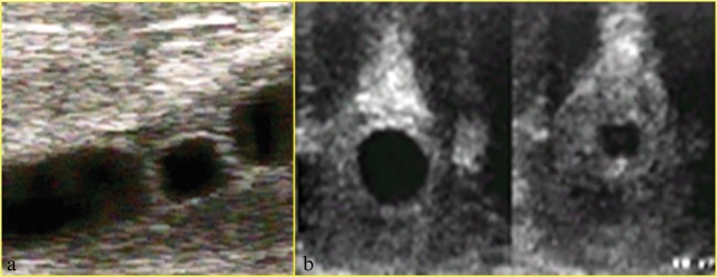
Viral papilloma seen in (a) longitudinal and (b) transverse planes.
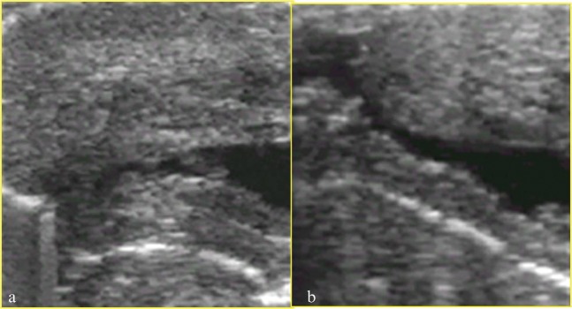
(a) Normal mucosa and periurethral tissues. (b) Periurethral fibrotic cuffing surrounding bulbar stricture.
Two examples of strictures within the fossa navicularis that could not be demonstrated on either contrast urethrography or ascending urethral ultrasound. The penile tip is to the left of the images.
Descending urethral ultrasound in hypospadias assessment demonstrating (a) a pinhole ventrally placed meatus and (b) a distal post-operative irregularity. The penile tip is to the left of the images.
Two attempts at performing descending urethral ultrasound in the same patient. (a) At the first attempt there was insufficient distension of the urethra. (b) This was rectified at the second attempt.
(a) Descending urethral ultrasound that fails to demonstrate a proximal stricture, which is identified on (b) perineal voiding images.
Similar articles
-
Sonography in the evaluation of the male anterior urethra.Clin Radiol. 1994 Sep;49(9):621-6. doi: 10.1016/s0009-9260(05)81879-3. Clin Radiol. 1994. PMID: 7955889
-
Imaging of the male urethra.Semin Ultrasound CT MR. 2007 Aug;28(4):258-73. doi: 10.1053/j.sult.2007.05.003. Semin Ultrasound CT MR. 2007. PMID: 17874650 Review.
-
[Ultrasonography in syringocele of the male urethra (ultrasound-urethrography)].Radiol Med. 1989 Oct;78(4):348-50. Radiol Med. 1989. PMID: 2687965 Italian.
-
Contrast-Enhanced Ultrasound: A Real-Time, Noninvasive, Radiation-Free Method for Intraoperative Male Urethral Fistula Assessment.J Ultrasound Med. 2024 Oct;43(10):1911-1918. doi: 10.1002/jum.16525. Epub 2024 Aug 3. J Ultrasound Med. 2024. PMID: 39096110
-
Ultrasonographic assessment of male anterior urethra. Description of the technique of examination and presentation of major pathologies.Med Ultrason. 2020 May 11;22(2):236-242. doi: 10.11152/mu-2426. Med Ultrason. 2020. PMID: 32399530 Review.
Cited by
-
Ultrasound imaging of male urethral stricture disease: a narrative review of the available evidence, focusing on selected prospective studies.World J Urol. 2024 Jan 13;42(1):32. doi: 10.1007/s00345-023-04760-x. World J Urol. 2024. PMID: 38217706 Free PMC article. Review.
-
Imaging the acute complications of gender-affirming surgeries: a primer for radiologists in the emergency setting.Abdom Radiol (NY). 2024 Aug;49(8):2812-2832. doi: 10.1007/s00261-024-04385-7. Epub 2024 Jun 4. Abdom Radiol (NY). 2024. PMID: 38832942 Review.
-
Three-Dimensional Imaging of Urethral Stricture Disease and Urethral Pathology for Operative Planning.Curr Urol Rep. 2016 Aug;17(8):54. doi: 10.1007/s11934-016-0616-0. Curr Urol Rep. 2016. PMID: 27278565 Review.
-
Male urogenital disorders.Br J Radiol. 2012 Nov;85 Spec No 1(Spec Iss 1):S1-2. doi: 10.1259/bjr/23337502. Br J Radiol. 2012. PMID: 23118098 Free PMC article. No abstract available.
-
Sonography of the distal urethra in lambs.Acta Vet Scand. 2017 Mar 14;59(1):16. doi: 10.1186/s13028-017-0283-2. Acta Vet Scand. 2017. PMID: 28292311 Free PMC article.
References
-
- McCallum RW. The adult male urethra: normal anatomy, pathology, and method of urethrography. Radiol Clin North Am 1979;17:227–44 - PubMed
-
- McAninch JW, Laing FC, Jeffrey Jr RB. 226. Sonourethrography in the evaluation of urethral strictures: a preliminary report. J Urol 1988;139:294–7 - PubMed
-
- Merkle W, Wagner W. Sonography of the distal male urethra—a new diagnostic procedure for urethral strictures: results of a retrospective study. J Urol 1988;140:1409–11 - PubMed
-
- Klosterman PW, Laing FC, McAninch JW. Sonourethrography in the evaluation of urethral stricture disease. Urol Clin North Am 1989;16:791–7 - PubMed
-
- Chiou RK, Anderson JC, Tran T, Patterson RH, Wobig R, Taylor RJ. Evaluation of urethral strictures and associated abnormalities using high-resolution and color Doppler ultrasound. Urology 1996;47:102–7 - PubMed
MeSH terms
LinkOut - more resources
Full Text Sources
Medical
