

doi: 10.1002/anie.201202204.
Epub 2012 Jun 5.
Re-engineering electrochemical biosensors to narrow or extend their useful dynamic range
Affiliations
- PMID: 22674785
- PMCID: PMC3482547
- DOI: 10.1002/anie.201202204
Item in Clipboard
Re-engineering electrochemical biosensors to narrow or extend their useful dynamic range
Angew Chem Int Ed Engl.
.
Abstract
Here we demonstrate two convenient methods to extend and narrow the useful dynamic range of a model electrochemical DNA sensor. We did so by combining DNA probes of different target affinities but with similar specificity on the same electrode. We were able to achieve an extended dynamic response spanning 3 orders of magnitude in target concentration. Using a different strategy we have also narrowed the useful dynamic range of an E-DNA sensor to only an 8-fold range of target concentrations.
Figures
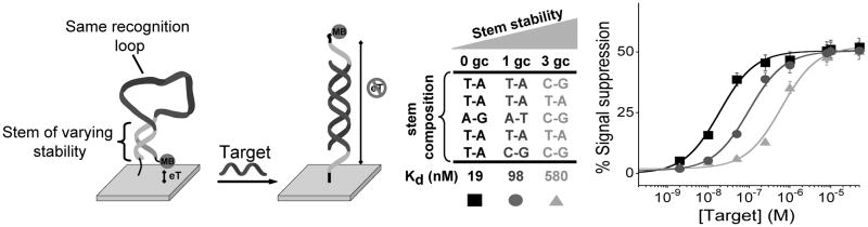
(Left) E-DNA sensors consist of a stem-loop DNA modified with a redox reporter (here methylene blue) and attached to an interrogating gold electrode via an introduced thiol group.[7b] This probe undergoes a large-scale conformational switch upon hybridization with a DNA complementary to the loop, leading to large change in Faradaic current from the redox reporter. The affinity of such “switch-based” probes can be rationally tuned by many orders of magnitude, without affecting their specificity, by simply altering the stability of their nonbinding, non-signalling state (e.g., by varying the stability of the E-DNA probe’s stem with the change of the GC base pairs content).[9] (Right) Here we have employed a set of three E-DNA probes sharing a common recognition element but spanning almost three orders of magnitude of target affinity. Error bars in this figure and in the following figures represent the average and standard deviations of measurements performed on at least three independently sensors.
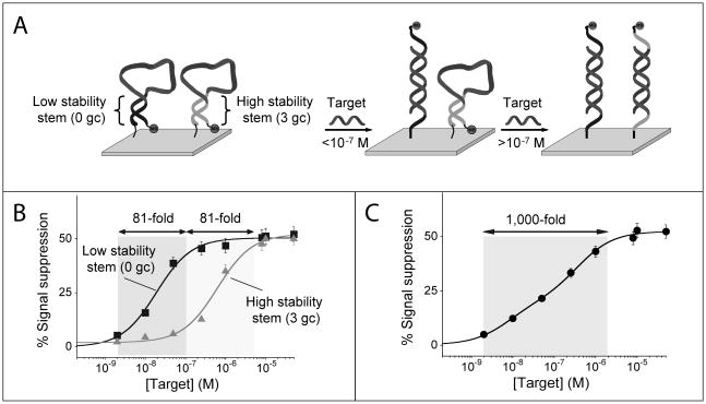
Employing a pair of signalling probes differing in affinity we can broaden the dynamic range of E-DNA sensors. (A) We did so by co-immobilizing (1:1 ratio) on a single electrode surface a relatively low affinity E-DNA probe (e.g., probe 3GC, Kd = 580 nM) with a higher affinity E-DNA probe (e.g., probe 0GC, Kd = 19 nM). (B) The useful dynamic range (defined as the fold-concentration change upon transition from 10% occupancy to 90% occupancy) of these individual probes spans an 81-fold range of target concentrations over two distinct concentration regimes. (C) With this strategy the resulting dose-response curve is extended and spans a 1,000-fold range of target concentrations.
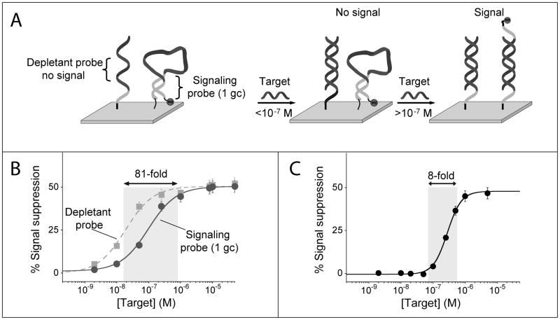
Using the sequestration mechanism we can dramatically narrow the useful dynamic range of an E-DNA sensor, thus greatly improving its sensitivity (i.e., its ability to measure small changes in target concentration). (A) We do so by co-immobilizing on a single electrode surface a low affinity, signaling E-DNA probe with a higher affinity probe (depletant) which, lacking the redox reporter, does not signal upon binding its target. At low concentrations the target preferentially binds the depletant, which removes (sequesters) target from the sample without generating a signal. When the total target amount surpasses that of the depletant (the sink is saturated), a threshold response is achieved in which further addition of target dramatically raises the relative concentration of free target. This gives rise to a much steeper dose-response curve than this would occur in the absence of a depletant. (C) Using this approach we have narrowed the 81-fold useful dynamic range of an unmodified E-DNA sensor to a mere 8-fold, thus increasing its sensitivity by an order of magnitude.
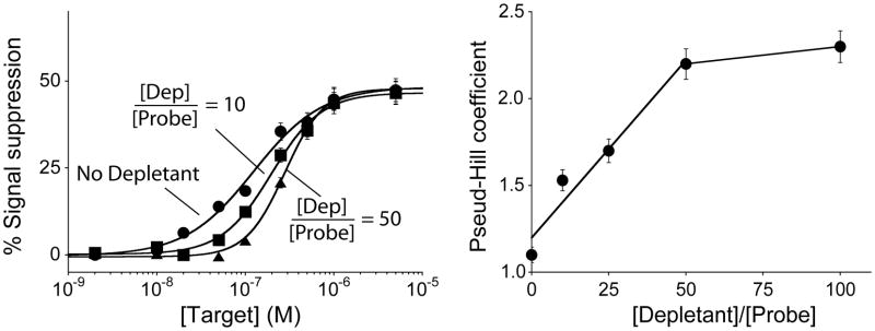
The sensitivity (i.e., steepness of the dose-response curve) achieved using the sequestration mechanism depends on the ratio of depletant to probe employed during sensor fabrication. To show this we have fitted our data to obtain pseudo-Hill coefficients, which, although our system is not classically cooperative, are analogous to the Hill coefficient commonly used to describe cooperative enzymatic systems.[13] We find that the pseudo-Hill coefficient increases monotonically with this ratio until plateauing at values above 50.
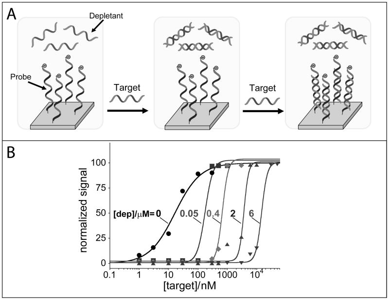
To overcome the limitations inherent to the surface attached depletants (which are easily saturated), we also show that the depletant probe can be simply added in solution at a fixed concentration. Here we use an unlabeled non-signalling probe (with the exact same sequence of the signalling redox-labelled probe) that sequesters the target DNA till a threshold level (fixed by the depletant concentration in solution) over which further increase in target concentration results in a steep dose-response curve. Because the depletant is free in solution, it rapidly reacts with the target (and with higher affinity) before this later can diffuse to the electrode surface and “activate” the signalling probe. (B) By using different concentrations of depletant in the reaction mix (0, 0.05, 0.4, 2, 6 μM) we can not only achieve steeper transitions than those observed with the depletant co-immobilized with the probe but we can also easily tune the threshold level at which we observe the sharp digital-like response of the sensor.
References
-
- Bullen RA, Arnot TC, Lakeman JB, Walsh FC. Biosens Bioelectron. 2006;21:2015–2045. - PubMed
-
- Privman V, Pedrosa V, Melnikov D, Pita M, Simonian A, Katz E. Biosens Bioelectron. 2009;25:695–701. - PubMed
- Wang J, Katz E. Isr J Chem. 2011;51:141–150.
-
- Koshland DE. The molecular basis for enzyme regulation. Vol. 1. Academic Press; New York: 1970.
- Goldbeter A, Koshland DE. Proc Natl Acad Sci USA. 1981;78:6840–6844. - PMC - PubMed
- Koshland DE, Goldbeter A, Stock JB. Science. 1982;217:220–225. - PubMed
- Ferrell JE. Trends Biochem Sci. 1996;21:460–466. - PubMed
Publication types
MeSH terms
Substances
Grants and funding
LinkOut - more resources
Full Text Sources
Other Literature Sources
