. 2012 Jul;29(7):275-91.
doi: 10.1002/yea.2908. Epub 2012 Jun 6.

A new method to efficiently induce a site-specific double-strand break in the fission yeast Schizosaccharomyces pombe

Affiliations

A new method to efficiently induce a site-specific double-strand break in the fission yeast Schizosaccharomyces pombe

Sham Sunder et al. Yeast. 2012 Jul.

Abstract

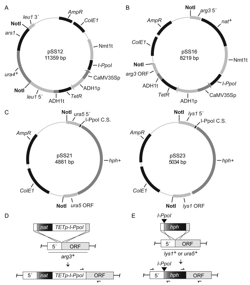
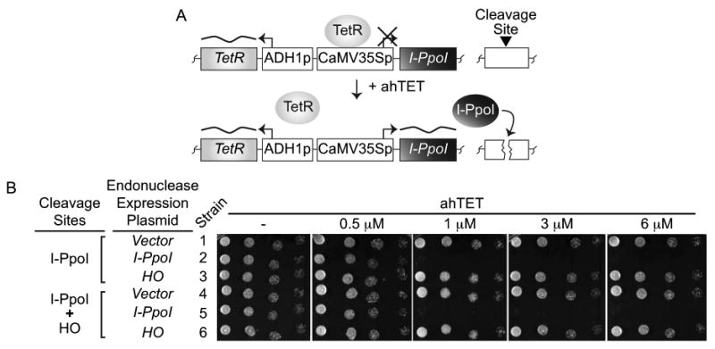
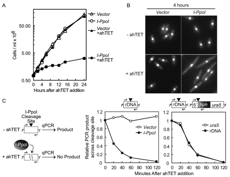
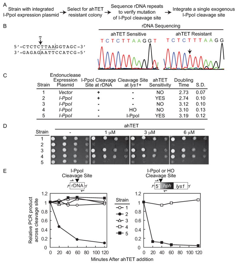
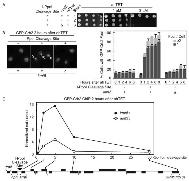
Double-strand DNA breaks are a serious threat to cellular viability and yeast systems have proved invaluable in helping to understand how these potentially toxic lesions are sensed and repaired. An important method to study the processing of DNA breaks in the budding yeast Saccharomyces cerevisiae is to introduce a unique double-strand break into the genome by regulating the expression of the site-specific HO endonuclease with a galactose inducible promoter. Variations of the HO site-specific DSB assay have been adapted to many organisms, but the methodology has seen only limited use in the fission yeast Schizosaccharomyces pombe because of the lack of a promoter capable of inducing endonuclease expression on a relatively short time scale (~1 h). We have overcome this limitation by developing a new assay in which expression of the homing endonuclease I-PpoI is tightly regulated with a tetracycline-inducible promoter. We show that induction of the I-PpoI endonuclease produces rapid cutting of a defined cleavage site (> 80% after 1 h), efficient cell cycle arrest and significant accumulation of the checkpoint protein Crb2 at break-adjacent regions in a manner that is analogous to published findings with DSBs produced by an acute exposure to ionizing irradiation. This assay provides an important new tool for the fission yeast community and, because many aspects of mammalian chromatin organization have been well-conserved in Sz. pombe but not in S. cerevisiae, also offers an attractive system to decipher the role of chromatin structure in modulating the repair of double-stranded DNA breaks.


Figures

Figure 1
Figure 1
Plasmids and integration strategies. Tetracycline inducible I-PpoI integration plasmids are shown in (A) and (B), I-PpoI cleavage site integration plasmids in (C), and integration strategies in (D) and (E) (not drawn to scale). (A) The I-PpoI ORF was cloned under the control of the CaMV35S promoter (p) and the nmt1 terminator (t) in the pDUAL-tet-rpsL-neo vector (Erler et al., 2006) to create pSS12. The plasmid also contains a tetracycline repressor (TetR) that is constitutively expressed from an ADH1 promoter and terminator. The TETp-I-PpoI NotI fragment from pSS12 (sites shown) is excised and integrated at leu1-32 as detailed (Matsuyama et al., 2004). (B) A second I-PpoI expression plasmid was created by transferring the entire TETp-I-PpoI control unit from pSS12 into a plasmid such that it was flanked by a clonNAT selectable marker (nat+) and sequences homologous to the regions immediately up (5′) and downstream (ORF) of the arg3+ ATG start codon. The TETp-I-PpoI NotI fragment is excised and integrated as shown in (D). PCR primers (355/512 Table I) used to verify correct integration at arg3+ are illustrated as half arrows. (C) To integrate an I-PpoI cleavage site at ura5+, pSS21 was created such that a single cleavage site was flanked by a hygromycin B selectable marker (hph+) marker and sequences homologous to the regions immediately up (5′) and downstream (ORF) of the ura5+ ATG start codon. A NotI fragment with the cleavage site (black triangle) is then excised and integrated at ura5+ as shown in (E). PCR primers used to verify correct integration across each junction (396/611 and 397/612 Tabel I) are illustrated. The pSS23 plasmid was created in a similar fashion for integration of a single I-PpoI cleavage site immediately upstream of the lys1+ ATG start codon (E). Verification primers for lys1+ integration correspond to 396/844 and 397/356 (Table I). For (B) and (C) plasmids, integrations produce an auxotrophic phenotype for the corresponding amino acid that can be used as a second means to verify correct integration. See Material and methods for further details.
Figure 2
Figure 2
A tetracycline inducible I-PpoI expression system. (A) Assay schematic. The homing endonuclease I-PpoI is under the control of a tetracycline inducible CaMV35S promoter (p) and an ADH1 promoter drives constitutive expression of the tetracycline repressor (TetR) that normally represses I-PpoI expression. Addition of anhydrotetracycline (ahTET) to growth media triggers induction of I-PpoI expression and cutting at endogenous I-PpoI cleavage sites (black triangle) located in the rDNA repeats or at a single exogenous cleavage site integrated elsewhere. (B) Induction of I-PpoI expression with ahTET produces efficient killing of S. pombe cells. Either empty vector or plasmids containing tetracycline inducible I-PpoI or HO alleles were integrated into strains containing endogenous rDNA I-PpoI cleavage sites with or without a single exogenous HO cleavage site integrated at lys1+. Serial dilutions of cells (1:5) were spotted onto EMMG agar plates containing the indicated amount of ahTET and grown at 30°C. See materials and methods for further details. Strains: 1-YSS154; 2-YSS151; 3-YSLS798; 4-YSLS802; 5-YSLS803; 6-YSLS804.
Figure 3
Figure 3
Induction of I-PpoI expression efficiently inhibits cell growth and produces rapid DSB formation. (A) Growth curve after I-PpoI induction. Strains with either an integrated empty vector (YSS154) or a tetracycline inducible I-PpoI allele (YSS151) were grown in EMMG with (+) or without 3 μM ahTET and growth was monitored by cell counting. Time across the x-axis denotes hours after ahTET addition. (B) I-PpoI expression produces cell elongation. Cells at the 4 hour time point in A were fixed in methanol and visualized by DAPI staining. Images have been intentionally overexposed to show the entire cell body. (C) Rapid cutting of endogenous and exogenous I-PpoI cleavage sites. Left, illustration of quantitative real-time PCR (qPCR) I-PpoI cutting assay, half arrows denote oligos that span the I-PpoI cleavage site (black triangle). See text for further details. Middle panel, rapid I-PpoI cleavage at the rDNA repeats. Strains with either integrated empty vector (YSS154) or a tetracycline inducible I-PpoI allele (YSS151) were treated with 3 μM ahTET and cell aliquots taken at the time points indicated after induction. Genomic DNA was prepared and qPCR used to monitor product formation across the I-PpoI cleavage sites located in the rDNA repeats (illustrated top). qPCR product formation at the uncut lys1+ locus was used as a normalization control. See Materials and methods for further details. Right panel, rapid I-PpoI cutting at both a single exogenous I-PpoI cleavage site and at the rDNA repeats. I-PpoI expression was induced in strain YSS226 containing both endogenous rDNA PpoI cleavage sites and a single cleavage site integrated at ura5+ (illustrated top). qPCR was then used to monitor product formation across both cleavage sites as described above.
Figure 4
Figure 4
Isolation of rDNA mutations resistant to I-PpoI cleavage. (A) Strategy used to produce strains containing I-PpoI resistant rDNA repeats and a single exogenous I-PpoI cleavage site. (B) Sequence of I-PpoI resistant rDNA repeats. Left, I-PpoI rDNA cleavage site with T insertion of resistant strains indicated. Right, typical DNA sequencing chromatograms of rDNA repeats from ahTET sensitive and resistant strains (strains 2 and 3 in C, respectively) with the T insertion site indicated by the arrow. (C) Characteristics of relevant strains. The procedure detailed in A was used to generate strain 3 (YSLS790-3) containing mutated rDNA repeats resistant to I-PpoI cutting from the parental I-PpoI expressing strain 2 (YSS151). Either a single HO (strain 4, YSLS793) or I-PpoI (strain 5, YSLS792) cleavage site was then integrated back into the genome at lys1+(illustrated in right panel of (E)). Strain 1 (YSS154) with an integrated empty vector was assayed as a control. Strains 3, 4, and 5 are all derived from strain 2. Doubling times were determined at 30°C in EMMG in the absence of ahTET and averaged from at least 3 independent experiments with the standard deviation (S.D.) shown. (D) Integration of a single exogenous I-PpoI cleavage site back into the genome of a strain with I-PpoI resistant rDNA repeats restores ahTET sensitivity. Spot tests were performed as described for Figure 2 with strains detailed in (C). (E) I-PpoI rapidly cleaves a single exogenous cleavage site but not mutated rDNA repeats. I-PpoI cutting assays were performed as detailed for Figure 3C using strains detailed in (C) and oligos that span either the I-PpoI cleavage sites in the rDNA repeats (left) or a single exogenous HO or I-PpoI cleavage site integrated at lys1+ (right). Cleavage sites are denoted by the black triangle. Note that data is from a single experiment where all five strains were processed simultaneously so that the rate of I-PpoI cutting observed at the rDNA and lys1+ cleavage sites is directly comparable.
Figure 5
Figure 5
The checkpoint protein Crb2 efficiently accumulates at an I-PpoI induced break in a manner that requires the H4K20 methylase Kmt5. (A) ahTET sensitivity of relevant strains. Strain 1 (YSS271) with an integrated tetracycline inducible I-PpoI allele, I-PpoI resistant rDNA repeats and GFP tagged crb2+ was generated and a single exogenous I-PpoI cleavage site then integrated at ura5+ (illustrated in (C)). to produce strain 2 (YSS289). The Kmt5 ORF was then replaced with a bsdMx6 marker to create Δkmt5 strain 3 (YSLS783). Spot tests were performed as detailed for Figure 2. (B) Live cell microscopy of GFP-Crb2 after induction of I-PpoI expression. The ahTET inducer was added to growing cultures of strains detailed in (A) to induce I-PpoI expression and live cell microscopy was performed at times indicated after ahTET addition. Left, representative images 2 hours after ahTET addition. Note that the nucleus of 3 different cells is denoted in each panel by GFP-Crb2 staining which is entirely nuclear. Arrowheads in middle panel denote GFP-Crb2 foci. Right, quantification of GFP-Crb2 foci, numbers were averaged from at least 3 independent experiments with ≥ 200 cells counted for each point. (C) ChIP of GFP-Crb2 at an I-PpoI induced break. Strains 2 and 3 from A were processed for anti-GFP ChIP 2 hours after induction of I-PpoI expression as described in Materials and methods. The relative enrichment of GFP-Crb2 at the break site was calculated by normalizing the cut/uncut ratio from each oligo pair flanking the cleavage site versus the cut/uncut ratio for an oligo pair at uncut lys1+. The location of the integrated I-PpoI cleavage site (black triangle), the hygromycin B selection mark (hph) and the approximate position of each oligo pair in the ura5-SPBC725.04 region of chromosome II is shown (drawn to scale, black boxes denote annotated ORFs, illustrated in the reverse complement orientation).

Similar articles

Cited by

References

    1. Ampatzidou E, Irmisch A, O'Connell MJ, Murray JM. Smc5/6 is required for repair at collapsed replication forks. Mol Cell Biol. 2006;26:9387–9401. - PMC - PubMed
    1. Bahler J, Wu JQ, Longtine MS, et al. Heterologous modules for efficient and versatile PCR-based gene targeting in Schizosaccharomyces pombe. Yeast. 1998;14:943–951. - PubMed
    1. Bannister AJ, Kouzarides T. Regulation of chromatin by histone modifications. Cell Res. 2011;21:381–395. - PMC - PubMed
    1. Bellaiche Y, Mogila V, Perrimon N. I-SceI endonuclease, a new tool for studying DNA double-strand break repair mechanisms in Drosophila. Genetics. 1999;152:1037–1044. - PMC - PubMed
    1. Berkovich E, Monnat RJJ, Kastan MB. Roles of ATM and NBS1 in chromatin structure modulation and DNA double-strand break repair. Nat Cell Biol. 2007;9:683–690. - PubMed

Publication types

MeSH terms

Substances

LinkOut - more resources