

Differential distribution patterns from medial prefrontal cortex and dorsal raphe to the locus coeruleus in rats
- PMID: 22674904
- PMCID: PMC3408042
- DOI: 10.1002/ar.22505
Differential distribution patterns from medial prefrontal cortex and dorsal raphe to the locus coeruleus in rats
Abstract
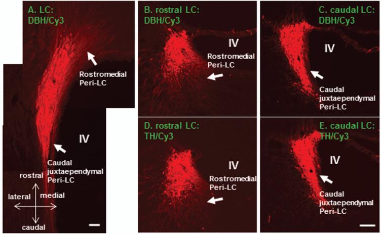
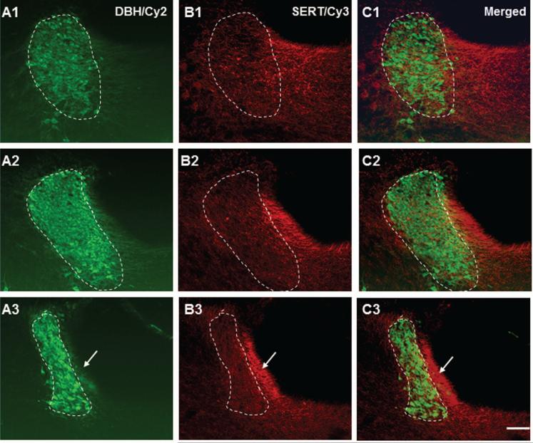
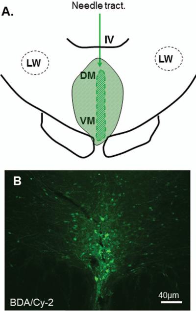
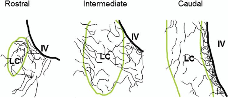
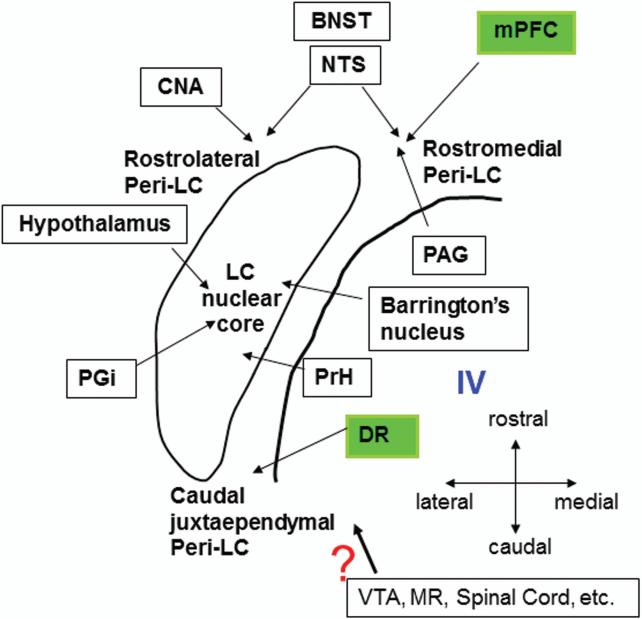
Locus coeruleus (LC) consists of a densely packed nuclear core and a surrounding plexus of dendritic zone, which is further divided into several subregions. Whereas many limbic-related structures topographically target specific subregions of the LC, the precise projections from two limbic areas, that is, medial prefrontal cortex (mPFC) and dorsal raphe (DR), have not been investigated. The goal of the present study is to identify and compare the distribution patterns of mPFC and DR afferent terminals to the LC nuclear core as opposed to specific pericoerulear dendritic regions (Peri-LC). To address these issues, anterograde tracer injections were combined with dopamine-β-hydroxylase (DBH) immunofluorescent staining to reveal the distribution patterns around the LC nuclear complex. Our data suggest that both mPFC-LC and DR-LC projections exhibit selective afferent terminal patterns. More specifically, mPFC-LC projecting fibers mainly target the rostromedial Peri-LC, whereas DR-LC projecting fibers demonstrate a preference to the caudal juxtaependymal Peri-LC. Thus, our present findings provide further evidences that afferents to the LC are topographically organized. Understanding the relationship among different inputs to the LC may help to elucidate the organizing principle which likely governs the interactions between the broad afferent sources of the LC and its global efferent targets.
Copyright © 2012 Wiley Periodicals, Inc.
Figures


Similar articles
-
Reciprocal connections between subdivisions of the dorsal raphe and the nuclear core of the locus coeruleus in the rat.Brain Res. 2004 Nov 5;1026(1):56-67. doi: 10.1016/j.brainres.2004.08.022. Brain Res. 2004. PMID: 15476697
-
Projections from melanin-concentrating hormone (MCH) neurons to the dorsal raphe or the nuclear core of the locus coeruleus in the rat.Brain Res. 2013 Jan 15;1490:72-82. doi: 10.1016/j.brainres.2012.08.022. Epub 2012 Sep 5. Brain Res. 2013. PMID: 22967922
-
Selective prefrontal cortical projections to the region of the locus coeruleus and raphe nuclei in the rhesus monkey.Brain Res. 1984 Jul 23;306(1-2):9-18. doi: 10.1016/0006-8993(84)90351-2. Brain Res. 1984. PMID: 6466989
-
Afferent regulation of locus coeruleus neurons: anatomy, physiology and pharmacology.Prog Brain Res. 1991;88:47-75. doi: 10.1016/s0079-6123(08)63799-1. Prog Brain Res. 1991. PMID: 1687622 Review.
-
Projection specificity in heterogeneous locus coeruleus cell populations: implications for learning and memory.Learn Mem. 2015 Sep 1;22(9):444-51. doi: 10.1101/lm.037283.114. Print 2015 Sep. Learn Mem. 2015. PMID: 26330494 Free PMC article. Review.
Cited by
-
The Noradrenergic System in Parkinson's Disease.Front Pharmacol. 2020 Apr 8;11:435. doi: 10.3389/fphar.2020.00435. eCollection 2020. Front Pharmacol. 2020. PMID: 32322208 Free PMC article. Review.
-
Resveratrol: A Multifaceted Guardian against Anxiety and Stress Disorders-An Overview of Experimental Evidence.Nutrients. 2024 Aug 26;16(17):2856. doi: 10.3390/nu16172856. Nutrients. 2024. PMID: 39275174 Free PMC article. Review.
-
After a period of forced abstinence, rats treated with the norepinephrine neurotoxin DSP-4 still exhibit preserved food-seeking behavior and prefrontal cortex fos-expressing neurons.Heliyon. 2024 Jun 4;10(13):e32146. doi: 10.1016/j.heliyon.2024.e32146. eCollection 2024 Jul 15. Heliyon. 2024. PMID: 39027623 Free PMC article.
-
Lasting neurobehavioral abnormalities in rats after neonatal activation of serotonin 1A and 1B receptors: possible mechanisms for serotonin dysfunction in autistic spectrum disorders.Psychopharmacology (Berl). 2014 Mar;231(6):1191-200. doi: 10.1007/s00213-013-3242-2. Epub 2013 Aug 24. Psychopharmacology (Berl). 2014. PMID: 23975037 Free PMC article.
-
Forebrain GABAergic projections to locus coeruleus in mouse.J Comp Neurol. 2013 Jul 1;521(10):2373-97. doi: 10.1002/cne.23291. J Comp Neurol. 2013. PMID: 23296594 Free PMC article.
References
-
- Amodio DM, Frith C. Meeting of minds: the medial frontal cortex and social cognition. Nat neurosci. 2006;7:268–277. - PubMed
-
- Amat J, Baratta MV, Paul E, Bland ST, Watkins LR, Maier SF. Medial prefrontal cortex determines how stressor controllability affect behavior and dorsal raphe nucleus. Nat Neurosci. 2005;8(3):365–371. - PubMed
-
- Arnsten AF, Goldman-Rakic PS. Selective prefrontal cortical projections to the region of the locus coeruleus and raphe nuclei in the Rhesus monkey. Brain Res. 1984;306:9–18. - PubMed
-
- Aston-Jones G, Ennis M, Pieribone VA, Nidkell VT, Shipley MT. The brain nucleus locus coeruleus: restricted afferent control of a broad efferent network. Science. 1986;234:734–737. - PubMed
Publication types
MeSH terms
Substances
Grants and funding
LinkOut - more resources
Full Text Sources
Miscellaneous
