. 2012 Aug;86(16):8634-44.
doi: 10.1128/JVI.00280-12. Epub 2012 Jun 6.

Infectious endogenous retroviruses in cats and emergence of recombinant viruses

Affiliations

Infectious endogenous retroviruses in cats and emergence of recombinant viruses

Yukari Anai et al. J Virol. 2012 Aug.

Abstract

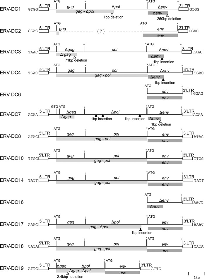
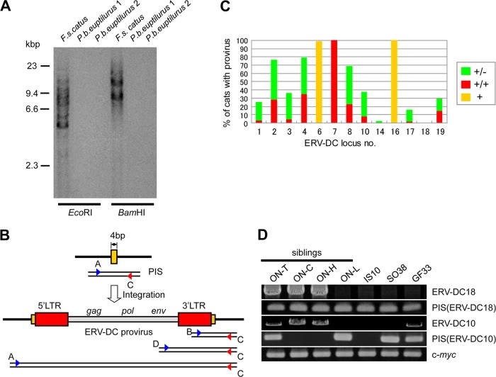
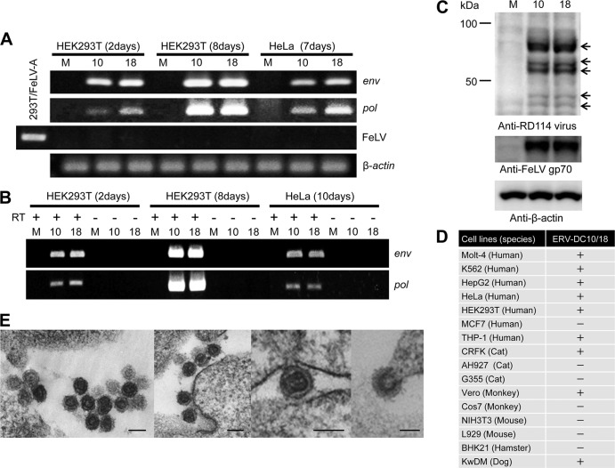
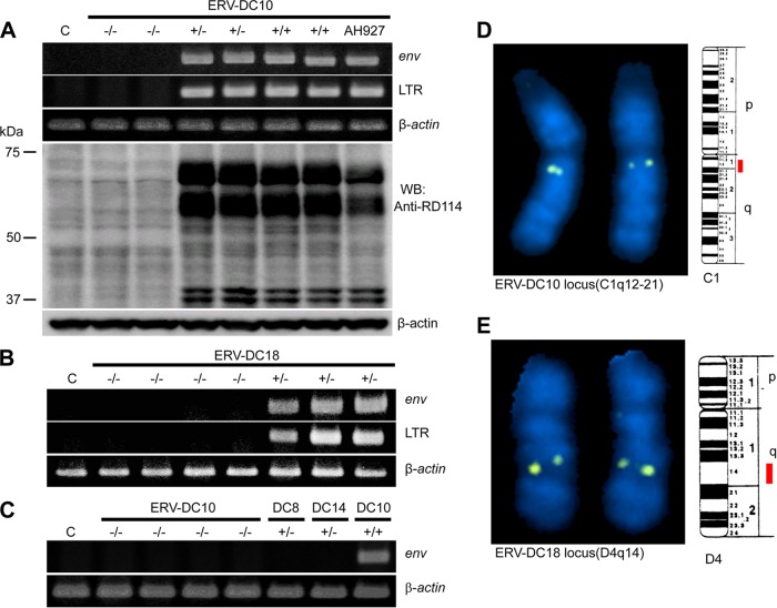
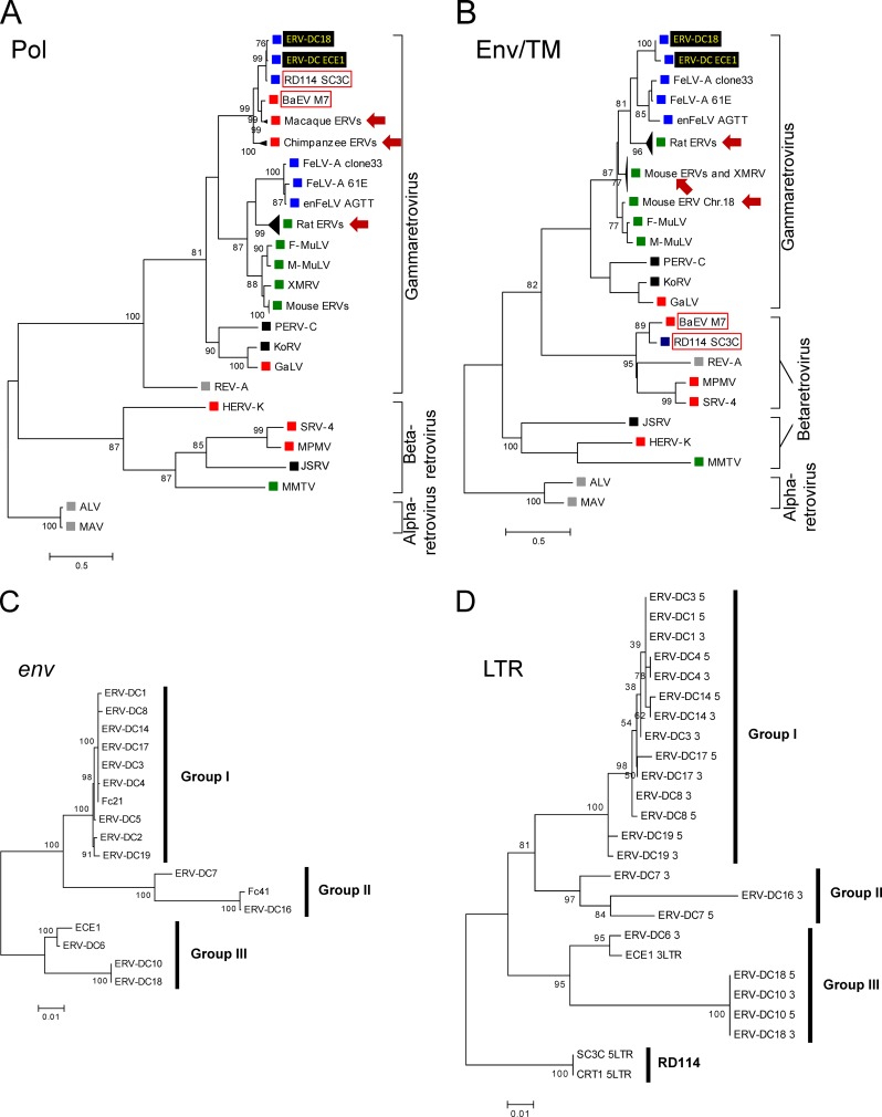
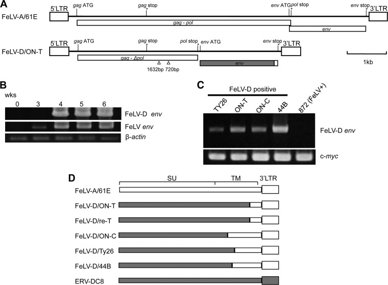
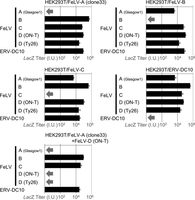
Endogenous retroviruses (ERVs) comprise a significant percentage of the mammalian genome, and it is poorly understood whether they will remain as inactive genomes or emerge as infectious retroviruses. Although several types of ERVs are present in domestic cats, infectious ERVs have not been demonstrated. Here, we report a previously uncharacterized class of endogenous gammaretroviruses, termed ERV-DCs, that is present and hereditary in the domestic cat genome. We have characterized a subset of ERV-DC proviral clones, which are numbered according to their genomic insertions. One of these, ERV-DC10, located in the q12-q21 region on chromosome C1, is an infectious gammaretrovirus capable of infecting a broad range of cells, including human. Our studies indicate that ERV-DC10 entered the genome of domestic cats in the recent past and appeared to translocate to or reintegrate at a distinct locus as infectious ERV-DC18. Insertional polymorphism analysis revealed that 92 of 244 domestic cats had ERV-DC10 on a homozygous or heterozygous locus. ERV-DC-like sequences were found in primate and rodent genomes, suggesting that these ERVs, and recombinant viruses such as RD-114 and BaEV, originated from an ancestor of ERV-DC. We also found that a novel recombinant virus, feline leukemia virus subgroup D (FeLV-D), was generated by ERV-DC env transduction into feline leukemia virus in domestic cats. Our results indicate that ERV-DCs behave as donors and/or acceptors in the generation of infectious, recombinant viruses. The presence of such infectious endogenous retroviruses, which could be harmful or beneficial to the host, may affect veterinary medicine and public health.


Figures

Fig 1
Fig 1
ERV-DC structures. Structures of the genomes of 10 full-length ERV-DC proviruses and three partial ERV-DC proviruses. The gag, pol, and env genes are illustrated together with the 5′ and 3′ LTRs and positions of the gag and env translational initiation codons (ATG). Asterisks indicate conserved stop codons. Gag and Pol proteins may be synthesized as a large single polypeptide precursor via termination suppression (44). An open triangle indicates a deletion of nucleotides, and a filled triangle indicates an insertion of nucleotides. Flanking 4-bp TSD sequences are shown for each provirus.
Fig 2
Fig 2
Detection of ERV-DC proviruses in the domestic cat genome. (A) Southern blotting of ERV-DC with an env probe derived from FeLV-D/ON-T in chromosomal DNA from the domestic cat (Felis silvestris catus) and Prionailurus bengalensis euptilurus. (B) PCR for detection of preintegration sites (PISs) and proviruses. Red boxes, viral LTRs; yellow boxes, target site duplications. Primers A to D are shown. The AC primer pair generated a PIS and/or full-length provirus. (C) Insertional polymorphisms of 13 ERV-DCs in 244 domestic cats. Green, provirus was detected on heterozygous loci; red, provirus was detected on homozygous loci; yellow, provirus was detected. (D) ERV-DC18 was only present in 3 of 4 siblings from one family. Each PIS, for full-length ERV-DC10 or ERV-DC18, was detected by primers Fe-122S and Fe-38R for ERV-DC10 or Fe-76S and Fe-81R for ERV-DC18 (see Table S4 in the supplemental material).
Fig 3
Fig 3
ERV-DC10 and ERV-DC18 are infectious viruses. (A) env and pol were detected by PCR at 2 and 8 days after infection of HEK293T cells. The supernatants from 8-day cultures were used to infect HeLa cells. M, mock infection; 10, infected with ERV-DC10; 18, infected with ERV-DC18. (B) Viruses collected from supernatants were used to detect env and pol genes by RT-PCR. (C) ERV-DC10 and ERV-DC18 proteins were detected in HEK293T cells using goat antiserum against disrupted RD-114 virions and anti-FeLV gp70 antibody. Anti-RD114 antibodies recognize many ERV-DC proteins (indicated by arrows) but not ERV-DC Env. (D) Susceptibility of cells to infection by ERV-DC10/ERV-DC18. (E) Transmission electron microscopy of retroviral particles in HEK293T cells persistently infected with ERV-DC10. The bar under each panel represents a 100-nm scale.
Fig 4
Fig 4
Active loci of ERV-DC10 and ERV-DC18. (A) ERV-DC10, (B) ERV-DC18, or (C) ERV-DC10, -DC8, or -DC14 DNA amplified by PCR with locus-specific primers using feline chromosomal DNA was directly transfected to HEK293T cells, and the filtrated supernatant was used to infect fresh HEK293T cells. Viral infection was monitored by detecting proviral DNAs or viral proteins. Each proviral genotype is indicated as −/− (wild), +/− (heterozygous), and +/+ (homozygous). Independent DNA samples were used in the figures. AH927 is a feline cell line with the ERV-DC10+/+ genotype. (D and E) FISH analysis of ERV-DC10 and ERV-DC18 loci on feline chromosomes.
Fig 5
Fig 5
Maximum-likelihood phylogenetic trees of Pol and Env TM proteins. We used amino acid sequences of entire regions of Pol proteins (A) and TM subunits of Env proteins (B) for 46 or 56 endogenous/exogenous viruses (see Table S5 in the supplemental material). A monophyletic subtree of the ERV elements obtained from the same species was compressed into a single branch, as indicated by arrows. Viral origin is indicated as blue (feline), orange (primate), green (rodent), gray (avian), or black (other species). The percent values were determined from 1,000 repeats of fast bootstrapping using RAxML (37) and are indicated at the branch junctions. More than 70 bootstrap values are shown. Maximum-likelihood phylogenic trees of ERV-DC env and LTR are indicated in panels C and D. Nucleotide sequences of entire ERV-DC env regions (C) or 5′ and 3′ LTRs in ERV-DCs (D) obtained were used. The general time-reversible model with rate heterogeneity among sites (GTR+Γ) was utilized. The percent values were determined from 1,000 repeats of fast bootstrapping using RAxML and are indicated at the branch junctions. Three or 5 indicates 3′ LTR or 5′ LTR. Sequences ECE1 (accession no. X51929), Fc21 (AF155060), Fc41 (AF155061), SC3C (EU030001), and CRT (AB559882) are indicated.
Fig 6
Fig 6
Transduction of ERV-DC env into FeLV. (A) Schematic representation of the structure of FeLV-D provirus integrated in spleen DNA of cat ON-T and FeLV-A/61E. White indicates FeLV sequences, and gray indicates ERV-DC sequences. (B) Isolation of FeLV-D in HEK293T cells inoculated with spleen extracts from cat ON-T. FeLV-D- and FeLV env-specific PCR was carried out. (C) A total of 283 DNA samples were screened for the presence of FeLV-D, and four positive samples were detected. (D) Structure of FeLV-D env. Recombination pattern of each FeLV-D env is indicated. White indicates FeLV sequences, and gray indicates ERV-DC sequences.
Fig 7
Fig 7
Infection assay of lacZ pseudotype virus. FeLV-A/Glasgow-1 env, FeLV-B/Gardner-Arnstein env, FeLV-C/Sarma env, FeLV-D/ON-T env, FeLV-D/Ty26 env, and ERV-DC10 env were used for each pseudotype virus preparation. HEK293T cells infected with FeLV-A/clone33, FeLV-B/Gardner-Arnstein, FeLV-C/Sarma, ERV-DC10, or both FeLV-A/clone33 and FeLV-D/ON-T were used as target cells for an interference assay. X-Gal-positive cells were counted as infectious units (I.U.). Arrow indicates viral interference.

Similar articles

Cited by

References

    1. Benveniste RE, Todaro GJ. 1974. Evolution of C-type viral genes: inheritance of exogenously acquired viral genes. Nature 252:456–459 - PubMed
    1. Benveniste RE, Sherr CJ, Todaro GJ. 1975. Evolution of type C viral genes: origin of feline leukemia virus. Science 190:886–888 - PubMed
    1. Beyer W, Mohring R, Drescher B, Notzel U, Rosenthal S. 1987. Molecular cloning of an endogenous cat retroviral element (ECE 1)–a recombinant between RD-114 and FeLV-related sequences. Brief report. Arch. Virol. 96:297–301 - PubMed
    1. Boeke JD, Stoye JP. 1997. Retrotransposons, endogenous retroviruses, and the evolution of retroelements, p 343–436 In Coffin JM, Hughes SH, Varmus HE. (ed), Retroviruses. Cold Spring Harbor Laboratory Press, Cold Spring Harbor, NY - PubMed
    1. Bonner TI, Todaro GJ. 1979. Carnivores have sequences in their cellular DNA distantly related to the primate endogenous virus, MAC-1. Virology 94:224–227 - PubMed

Publication types

Associated data

LinkOut - more resources