. 2012 Aug;86(16):8693-704.
doi: 10.1128/JVI.00252-12. Epub 2012 Jun 6.

Kaposi's sarcoma-associated herpesvirus ORF54/dUTPase downregulates a ligand for the NK activating receptor NKp44

Affiliations

Kaposi's sarcoma-associated herpesvirus ORF54/dUTPase downregulates a ligand for the NK activating receptor NKp44

Alexis Spain Madrid et al. J Virol. 2012 Aug.

Abstract

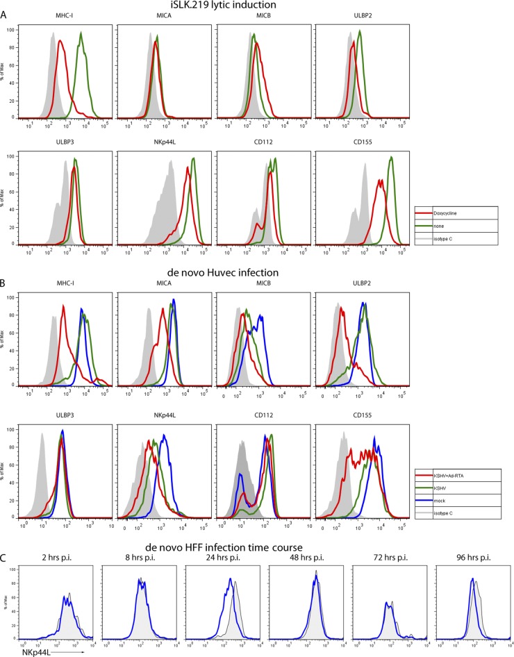
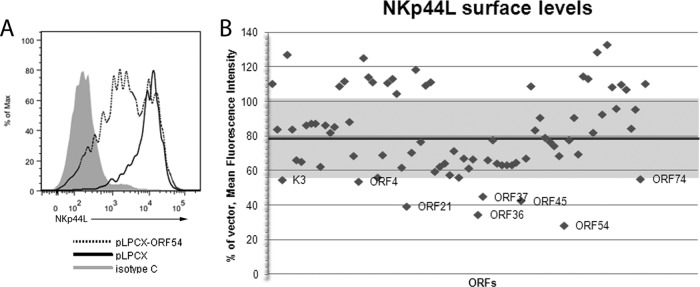
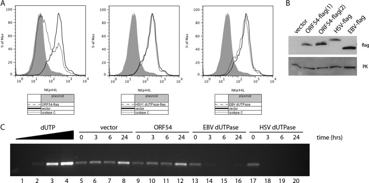
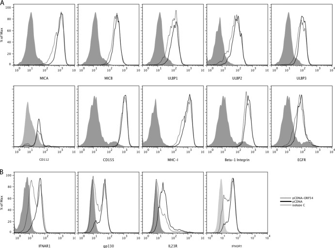
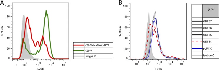
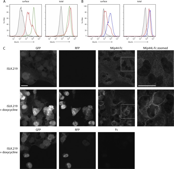
Kaposi's sarcoma-associated herpesvirus (KSHV) establishes long-term latent infection in humans and can cause cancers in endothelial and B cells. A functioning immune system is vital for restricting viral proliferation and preventing KSHV-dependent neoplasms. While natural killer (NK) lymphocytes are known to target virus-infected cells for destruction, their importance in the anti-KSHV immune response is not currently understood. Activating receptors on NK cells recognize ligands on target cells, including the uncharacterized ligand(s) for NKp44, termed NKp44L. Here we demonstrate that several NK ligands are affected when KSHV-infected cells are induced to enter the lytic program. We performed a screen of most of the known KSHV genes and found that the product of the ORF54 gene could downregulate NKp44L. The ORF54-encoded protein is a dUTPase; however, dUTPase activity is neither necessary nor sufficient for the downregulation of NKp44L. In addition, we find that ORF54 can also target proteins of the cytokine receptor family and the mechanism of downregulation involves perturbation of membrane protein trafficking. The ORF54-related proteins of other human herpesviruses do not possess this activity, suggesting that the KSHV homolog has evolved a novel immunoregulatory function and that the NKp44-NKp44L signaling pathway contributes to antiviral immunity.


Figures

Fig 1
Fig 1
NK activating ligand surface expression during KSHV infection. (A) Stably infected iSLK.219 cells were either left untreated (green lines) or treated with 1 μg/ml doxycycline (red lines) for 48 h to induce ectopic expression of the KSHV lytic switch protein RTA. Cells were stained with antibodies against NK activating ligands and analyzed by flow cytometry. Doxycycline-treated lytic cells denoted by the red line have been gated on the RFP-positive population. Isotype control (isotype C) staining is shown in gray-filled histograms. Doxycycline-induced RTA in cells without KSHV had no effects on any of the proteins tested (data not shown). (B) HUVECs were infected with KSHV (green lines), and half were induced to become lytic with Ad-RTA (red lines). These cells were compared to uninfected cells (blue lines) or cells treated with Ad-RTA alone (data not shown). Ad-RTA treatment on uninfected cells had no effects on the proteins tested, with the exception of MICB (data not shown). Isotype control staining is shown in gray-filled histograms. (C) HFF cells were infected with KSHV for the indicated times. Cells at each time point were harvested and stained with NKp44-Fc. Infected cells (blue lines) were compared to mock-infected cells (gray filled histograms). p.i., postinfection.
Fig 2
Fig 2
ORF54 was identified in a screen for NKp44L downregulation. A plasmid library containing most of the known KSHV genes was transfected individually into 293 cells together with a plasmid expressing GFP. At 48 h posttransfection, the cells were stained with NKp44-Fc and analyzed by flow cytometry. (A) Histogram comparing the NKp44L levels of vector alone (solid line) versus ORF54 (dotted line) in GFP-positive cells. Isotype control staining is shown in gray. (B) Dot plot depicting the NKp44L mean fluorescence intensity of the screened KSHV genes, including ORF54, relative to a vector control. The median value of 79 is depicted by the dark line, and the shaded area denotes 1 standard deviation above and below the median (SD = 24). The eight genes that fell below 1 SD in the primary screen are labeled.
Fig 3
Fig 3
NKp44L downregulation is independent of dUTPase activity. Flag-tagged versions of the dUTPases from KSHV, HSV-1, and EBV were transiently transfected into 293 cells. (A) At 48 h posttransfection, cells were stained with NKp44-Fc and analyzed by flow cytometry. Cells transfected with each dUTPase (dotted lines) were compared to empty vector control (solid lines). Isotype control staining is shown in gray-filled histograms. Results are representative of three individual experiments. (B) Lysates were prepared from the same cells used for panel A, equal amounts of total protein were loaded onto a gel, and the membrane was blotted with an anti-Flag antibody, as well as an anti-pyruvate kinase (PK) antibody as a loading control. (C) Flag-tagged dUTPases were immunoprecipitated from lysates and eluted with Flag peptide. dUTPase activity was measured by incubating this purified protein with dUTP for 0, 3, 6, or 24 h at 37°C. The resulting mixture was used in place of dTTP in a PCR to measure the amount of remaining dUTP in the reaction. As a control to show dUTP concentration dependence, various amounts of pure dUTP were added to the PCR mixture: 0 (lane 1), 6.25 μM (lane 2), 12.5 μM (lane 3), or 25 μM (lane 4). Time courses of dUTPase assays for vector control (lanes 5 to 8), ORF54 (lanes 9 to 12), EBV dUTPase (lanes 13 to 16), and HSV-1 dUTPase (lanes 17 to 20).
Fig 4
Fig 4
ORF54 specificity. 293 cells were transiently transfected with either ORF54 plasmid (dotted lines) or empty vector (solid lines) and stained with a panel of antibodies. (A) Flow cytometric cell surface staining of proteins that were unaffected by ORF54 expression (MICA, MICB, ULBP1, ULBP2, ULBP3, CD112, CD155, MHC-I, beta-1 integrin, and EGFR); (B) flow cytometric staining of proteins whose levels decreased upon ORF54 expression (IFNAR1, gp130, IL-23R, and IFNGR1).
Fig 5
Fig 5
IL-23R is downregulated by lytic infection and ORF54. (A) HFF cells stably infected with the recombinant rKSHV.219 were either left untreated (green line) or treated with 9 mM sodium butyrate (NaB) plus Ad-RTA (red line) for 72 h to induce lytic reactivation. Cells were stained with an antibody against IL-23R and analyzed by flow cytometry. Lytic cells denoted by the red line have been gated on the RFP-positive population. Isotype control staining is shown in gray-filled histograms. Uninfected HFF cells treated with Ad-RTA and/or NaB did not have altered IL-23R levels (data not shown). (B) A plasmid library containing most of the known KSHV genes was transfected individually into 293 cells together with a plasmid expressing GFP. At 48 h posttransfection, the cells were stained for IL-23R and analyzed by flow cytometry. Histogram comparing the IL-23R levels of vector alone (blue line) versus ORF54 (red dotted line) and several other representative ORFs in GFP-positive cells. Isotype control staining is shown in gray.
Fig 6
Fig 6
NKp44L subcellular localization is altered. (A) iSLK.219 cells were treated for 48 h with 1 μg/ml doxycycline to induce RTA expression and then stained with NKp44-Fc either without (left) or with (right) permeabilization. Latent cells are shown in green; lytic cells are in red. Isotype control staining is the gray-filled histogram. (B) 293 cells were transfected with vector control (blue line) or ORF54 (red line) and then stained with NKp44-Fc either without (left) or with (right) permeabilization. Isotype control staining is the gray-filled histogram. (C) iSLK.219 cells were grown without (row 1) or with (rows 2 and 3) 1 μg/ml doxycycline for 48 h and then fixed and stained with NKp44-Fc. Cells were imaged by confocal fluorescence microscopy. The boxed region of the NKp44L stain is shown blown up in the far right panel. Bars, 25 μm.

Similar articles

Cited by

References

    1. Ambroziak JA, et al. 1995. Herpes-like sequences in HIV-infected and uninfected Kaposi's sarcoma patients. Science 268:582–583 - PubMed
    1. Barabas O, Pongracz V, Kovari J, Wilmanns M, Vertessy BG. 2004. Structural insights into the catalytic mechanism of phosphate ester hydrolysis by dUTPase. J. Biol. Chem. 279:42907–42915 - PubMed
    1. Bechtel J, Grundhoff A, Ganem D. 2005. RNAs in the virion of Kaposi's sarcoma-associated herpesvirus. J. Virol. 79:10138–10146 - PMC - PubMed
    1. Bechtel JT, Liang Y, Hvidding J, Ganem D. 2003. Host range of Kaposi's sarcoma-associated herpesvirus in cultured cells. J. Virol. 77:6474–6481 - PMC - PubMed
    1. Bergman AC, Nyman PO, Larsson G. 1998. Kinetic properties and stereospecificity of the monomeric dUTPase from herpes simplex virus type 1. FEBS Lett. 441:327–330 - PubMed

Publication types

MeSH terms

LinkOut - more resources