. 2012 Aug;86(16):8740-9.
doi: 10.1128/JVI.07167-11. Epub 2012 Jun 6.

Hepatitis C virus infection alters P-body composition but is independent of P-body granules

Affiliations

Hepatitis C virus infection alters P-body composition but is independent of P-body granules

Gemma Pérez-Vilaró et al. J Virol. 2012 Aug.

Abstract

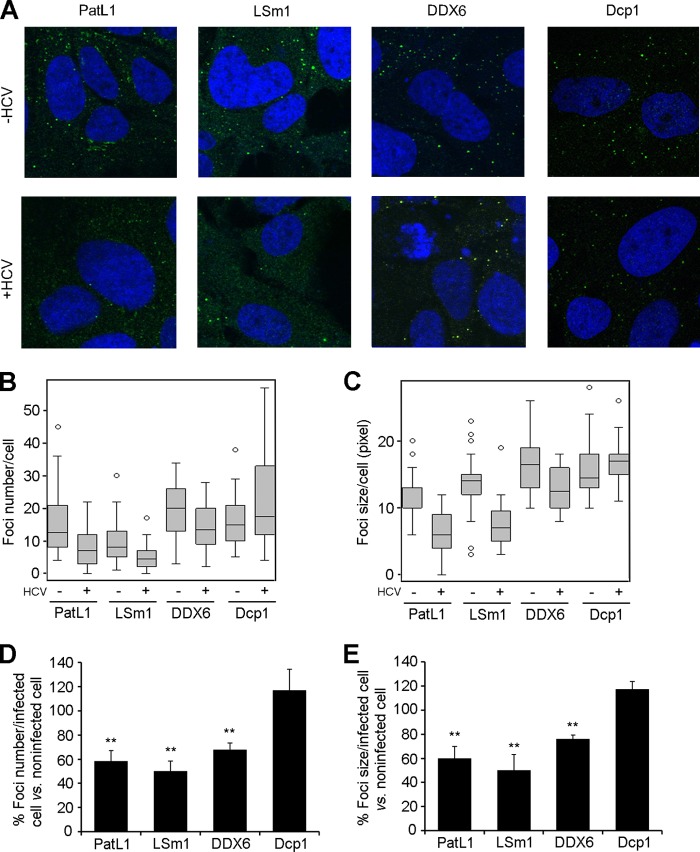
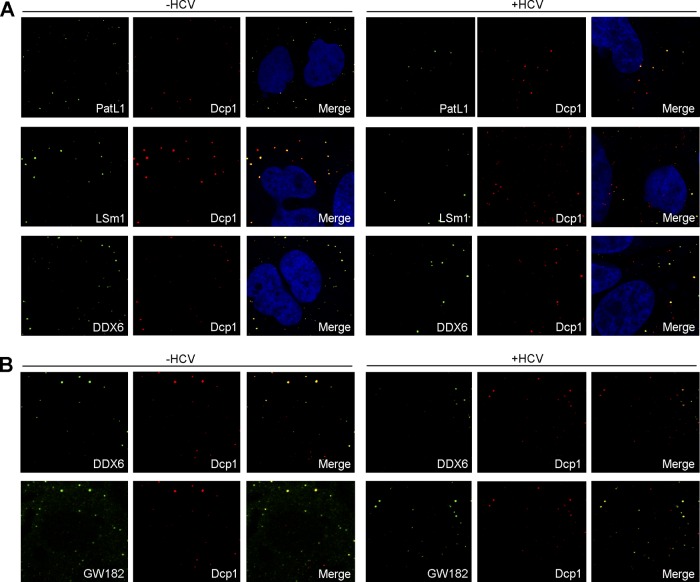
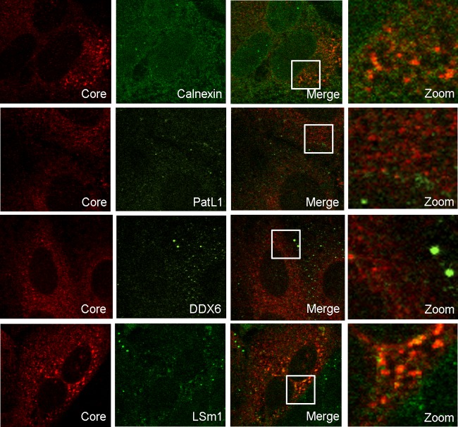
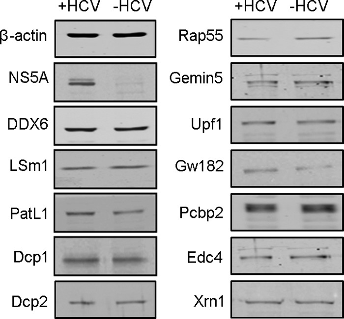
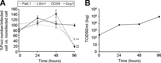
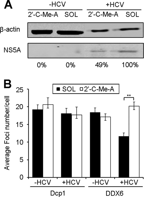
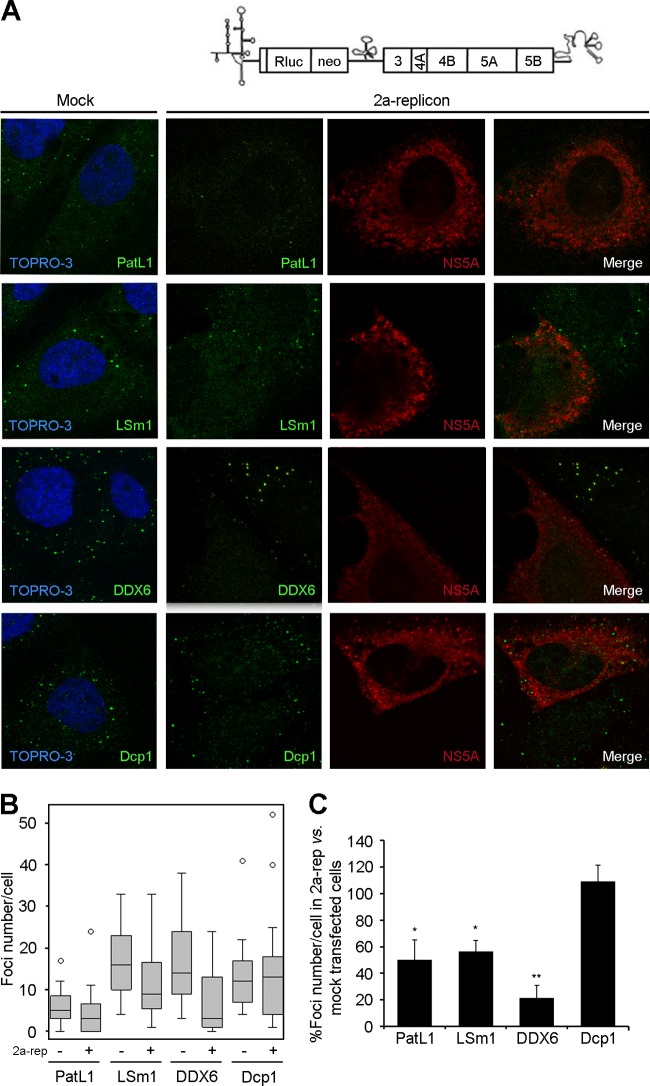
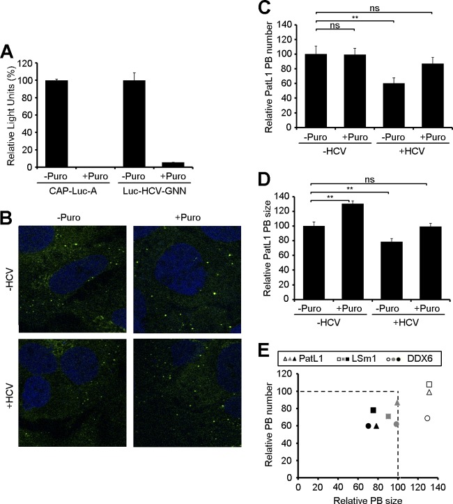
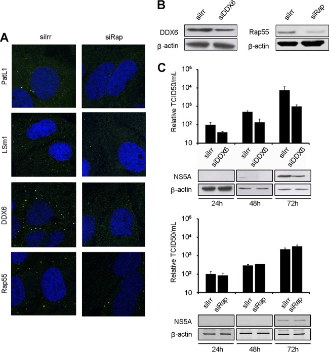
Processing bodies (P-bodies) are highly dynamic cytoplasmic granules conserved among eukaryotes. They are present under normal growth conditions and contain translationally repressed mRNAs together with proteins from the mRNA decay and microRNA (miRNA) machineries. We have previously shown that the core P-body components PatL1, LSm1, and DDX6 (Rck/p54) are required for hepatitis C virus (HCV) RNA replication; however, how HCV infection affects P-body granules and whether P-body granules per se influence the HCV life cycle remain unresolved issues. Here we show that HCV infection alters P-body composition by specifically changing the localization pattern of P-body components that are required for HCV replication. This effect was not related to an altered expression level of these components and could be reversed by inhibiting HCV replication with a polymerase inhibitor. Similar observations were obtained with a subgenomic replicon that supports only HCV translation and replication, indicating that these early steps of the HCV life cycle trigger the P-body alterations. Finally, P-body disruption by Rap55 depletion did not affect viral titers or HCV protein levels, demonstrating that the localization of PatL1, LSm1, and DDX6 in P-bodies is not required for their function on HCV. Thus, the HCV-induced changes on P-bodies are mechanistically linked to the function of specific P-body components in HCV RNA translation and replication; however, the formation of P-body granules is not required for HCV infection.


Figures

Fig 1
Fig 1
HCV infection alters P-body composition. (A) Huh7.5 cells were infected for 96 h with HCV and immunostained with antibodies for PatL1, LSm1, DDX6, or Dcp1 (green). Nuclei were visualized using DAPI or TOPRO-3 (blue). Z-series from 30 randomly selected cells under each condition were collected. (B and C) P-body number (B) and size (C) were quantified using the maximum-intensity projection image along the z axis. Graphed are box plots showing medians, upper and lower quartiles, and outliers. (D and E) Results for the number (D) and size (E) of foci containing PatL1, LSm1, DDX6, or Dcp1 in HCV-infected cells were also plotted as percentages relative to those in noninfected cells. Error bars indicate the standard error of the mean (**, P < 0.005).
Fig 2
Fig 2
Colocalization of different P-body markers after HCV infection. Huh7.5 cells were noninfected or infected with HCV. (A) After 96 h, cells were double immunostained with antibodies against Dcp1 (red) and PatL1, LSm1, or DDX6 (green) (B) or triple immunostained with antibodies against DDX6, GW182 (green), and Dcp1 (red) (B). Note that to clearly show colocalization patterns, the images were displayed as Dcp1-DDX6 and Dcp1-GW182 colocalization pairs. Images correspond to the same cells. Nuclei were visualized using DAPI (blue).
Fig 3
Fig 3
LSm1 colocalizes with the HCV core protein in a subset of HCV-infected cells. HCV-infected Huh7.5 cells were coimmunostained for HCV core protein and calnexin, PatL1, DDX6, or LSm1. Indicated areas in the merged images are magnified in the right column.
Fig 4
Fig 4
HCV infection does not affect the abundance of P-body components. Western blotting of core P-body components at 96 h after HCV infection is shown. NS5A and β-actin are shown as controls for infection and protein loading, respectively.
Fig 5
Fig 5
Kinetics of P-body abundance during HCV infection. (A) Huh7.5 cells were infected with HCV and at different times postinfection were immunostained to quantify PatL1-, LSm1-, DDX6-, and Dcp1-containing P-bodies in at least 100 cells. Shown are the relative abundances of PatL1-, LSm1-, and DDX6- versus Dcp1-containing P-bodies in HCV-infected relative to noninfected cells during the first 96 h postinfection. The asterisks indicate the time point at which differences were statistically significant (**, P < 0.005). (B) Kinetics of HCVcc infectivity in the supernatant of HCV-infected cells in a representative experiment during the first 96 h of infection. Error bars indicate the standard error of the mean. Note that the error bars in panel B are too small to be visible for some time points.
Fig 6
Fig 6
Treatment of HCV-infected cells with a polymerase inhibitor results in relocalization of DDX6 into P-bodies. Huh7.5 cells were infected with HCV for 84 h and treated at 48 h before sampling with 2′-C-methyladenosine (2′-C-Me-A), a nucleoside analog, or with the solvent (SOL) as a control. (A) NS5A protein levels were tested by Western blotting using β-actin as a control for protein loading. Numbers below the panel indicate the percentage of NS5A expression levels relative to those detected in solvent-treated cells. (B) Cells were immunostained as described for Fig. 1A, and P-body numbers in at least 100 cells were quantified. Error bars indicate the standard error of the mean (**, P < 0.005).
Fig 7
Fig 7
HCV RNA translation and replication alter P-body composition. (A) Huh7.5 cells were electroporated with a subgenomic 2a replicon RNA and coimmunostained after 48 h to detect NS5A and PatL1, LSm1, DDX6, or Dcp1. (B) Z-series from 30 randomly selected cells under each condition were collected. P-body number was quantified using the maximum-intensity projection image along the z axis. Graphed are box plots showing medians, upper and lower quartiles, and outliers. (C) Results for the number of foci containing PatL1, LSm1, DDX6, and Dcp1 in replicon-transfected NS5A-positive cells were also plotted as percentages relative to those in mock-transfected cells. Error bars indicate the standard error of the mean (*, P < 0.01; **, P < 0.005).
Fig 8
Fig 8
Effect of puromycin treatment on P-bodies in HCV-infected cells. (A) Huh7.5 cells were electroporated with a CAP-Luc-poly(A) derivative (CAP-Luc-A) or a nonreplicating HCVcc RNA that expresses luciferase protein (Luc-HCV-GNN). After electroporation, cells were treated for 1 h with 100 μg/ml puromycin, and luciferase activity was measured. (B) Noninfected and HCV-infected cells were treated at 96 h postinfection with 100 μg/ml of puromycin for 1 h and immunostained with antibodies against PatL1. (C and D) The number (C) and size (D) of PatL1-containing P-bodies (PB) from at least 100 cells were quantified and compared to those detected in noninfected, nontreated cells. Error bars indicate the standard error of the mean (*, P < 0.01; **, P < 0.005). (E) Similar results were obtained for DDX6- and LSm1-containing P-bodies, as shown in a composite figure. The number and size of P-bodies containing PatL1 (triangles), LSm1 (squares), or DDX6 (circles) from at least 100 cells were quantified. Shown are the average values for HCV-infected, puromycin-treated cells (gray), HCV-infected, nontreated cells (black), and noninfected, puromycin-treated cells (white) relative to those for noninfected, nontreated cells.
Fig 9
Fig 9
P-body disruption does not affect HCV infection. Huh7.5 cells were transfected with siRNA targeting Rap55 (siRap) or DDX6 (siDDX6) or with nontargeting siRNA (siIrr). (A) Immunostaining with antibodies against DDX6, LSm1, PatL1, or Rap55 (green). Nuclei were visualized using DAPI or TOPRO-3 (blue). (B) Immunoblot analysis of DDX6, Rap55, and β-actin. (C) DDX6 (upper panel)- and Rap55 (lower panel)-silenced cells were transfected with HCV RNA at the time of maximum silencing, and infectivity in the supernatant was monitored for 72 h in a limiting-dilution assay. Intracellular accumulation of HCV NS5A and cellular β-actin was monitored by immunoblotting (panels below graphs). Error bars indicate the standard error of the mean.

Similar articles

Cited by

References

    1. Aizer A, et al. 2008. The dynamics of mammalian P body transport, assembly, and disassembly in vivo. Mol. Biol. Cell 19:4154–4166 - PMC - PubMed
    1. Alves-Rodrigues I, Galao RP, Meyerhans A, Diez J. 2006. Saccharomyces cerevisiae: a useful model host to study fundamental biology of viral replication. Virus Res. 120:49–56 - PMC - PubMed
    1. Andrei MA, et al. 2005. A role for eIF4E and eIF4E-transporter in targeting mRNPs to mammalian processing bodies. RNA 11:717–727 - PMC - PubMed
    1. Angus AG, et al. 2010. Requirement of cellular DDX3 for hepatitis C virus replication is unrelated to its interaction with the viral core protein. J. Gen. Virol. 91:122–132 - PMC - PubMed
    1. Ariumi Y, et al. 2007. DDX3 DEAD-box RNA helicase is required for hepatitis C virus RNA replication. J. Virol. 81:13922–13926 - PMC - PubMed

Publication types

LinkOut - more resources